
Translate this page into:
Effect of mobile phone radiofrequency electromagnetic radiations on oxidative stress and feeding behaviour in Sprague Dawley (SD) rats
*Corresponding author: Pravallika Pagadala, Department of Physiology, Shridevi Institute of Medical Sciences and Research Hospital, Tumkuru, Karnataka, India. pravallikapagadala5@gmail.com
-
Received: ,
Accepted: ,
How to cite this article: Pagadala P, Shankar MSV, Sumathi M E. Effect of mobile phone radio frequency electromagnetic radiations on oxidative stress and feeding behaviour in Sprague Dawley (SD) rats. Indian J Physiol Pharmacol 2023;67:131-5.
Abstract
Objectives:
Radiofrequency electromagnetic radiation (RF-EMR) from mobile phones is known to produce a stress response because of its effect on hypothalamus. Mobile phones have become an integral part of our lives with increasing usage not only in terms of number of users but also increase in talk time. The present study aimed to study the effect of mobile phone radiofrequency electromagnetic radiations on oxidative stress and feeding behaviour assessment in Sprague Dawley (SD) rats.
Materials and Methods:
Twelve male SD rats of 10–12 weeks old, weighing 180–220 g, were housed and allowed to acclimatise in a room with 12:12 h light-dark cycle with ad libitum amount of food and reverse osmosis (RO) water before the start of the study. Then, rats were divided into control and RF-EMR exposed groups, and everyday feed intake and body weight were measured. At the end of the study period, blood sample was collected through retro orbital puncture for biochemical investigations.
Results:
The present study showed significant increase in malondialdehyde and serum corticosterone levels and decrease feeding behaviour in rats exposed to RF-EMR in rats exposed to RF-EMR.
Conclusion:
This study proves that mobile RF-EMR causes oxidative stress and oxidative damage leading to decreased feeding behaviour in SD rats.
Keywords
Oxidative damage
Reactive oxygen species
Neuronal cells
Brain damage
Body weight
Altered
Decibel meter

INTRODUCTION
With increased usage, not just in terms of the number of users, but also in terms of conversation time, mobile phones have become a vital part of our lives.[1] With the increased exposure to Radiofrequency emitted radiations (RF-EMR) and its effect on biological systems is a rising field of interest. Concerns regarding the potential health consequences of mobile phone use are growing as the number of users has exploded in recent years. Mobile phone technology makes use of RF-EMR and has greatly increased the amount of RF-EMR exposure people are exposed to on a daily basis. Much recent research has raised concerns about the safety of RF-EMR exposure in this way.[2] Many researchers also believe that the effects of mobile phone exposure may be due to the cumulative effects of the heat generated and the RF-EMR emitted from mobile phones. They have suggested that the contribution of the non-thermal component is minimal and that the effects of mobile phone exposure would be negligible if the thermal effect could be eliminated.[1]
Because of their effect on the hypothalamus, RF-EMRs from cell phones are known to create stress. Oxidative stress, which causes oxidative damage, is a major method by which stress affects the brain.[1] Oxidative stress occurs when there is a mismatch between the systemic expression of reactive oxygen species (ROS) and the biological system’s ability to quickly detoxify the reactive intermediates or repair the harm they cause during stressful situations. Disturbances in a cell’s natural condition can result in harmful effects from the formation of peroxides and free radicals, which can harm all of the cell’s components. It has been linked to increased generation of oxidising species and can create abnormalities in normal cellular signalling systems.[2] The effects of RF-EMR are also thought to be caused by oxidative stress, which is defined as the excessive production of ROS.[1] This behaviour has been observed in Drosophila whole body and ovarian tissue models, mouse fibroblasts, cultured breast cancer cells, rat heart tissue and human lens epithelial cells after RF-EMR treatment.[3-5] A stressful situation induces the organism to activate not only the adrenal system but also the central nervous system and the pituitary.[6,7] Stress is a challenge to the natural homeostasis of an organism; in turn, the organism may react to stress by producing a physiological response to regain equilibrium lost by the impact of the stressor.[8,9] When the stressor exceeds a organisms ability to withstand or ability to respond appropriately to the stress, the homeostasis is disturbed. One such homeostasis that is disrupted is in feeding behaviour.[10] Feeding behaviour is related to obtaining and consuming food.[11] It is a complex behaviour in our day to day life that is regulated by many mechanisms.[12-14] However, the exact regulatory mechanisms are not well understood.[11]
Studies showed that stress also causes oxidative damage leading to oxidative stress effect on brain cerebral blood flow, blood brain barrier and neuronal damage.[1,2]
Oxidative stress and lipid peroxidation are known by measuring parameters such as malondialdehyde (MDA) and serum corticosterone levels.[15-17] Thus, the present study aimed to study the effect of mobile phone RF-EMR on feeding behaviour, MDA and serum corticosterone levels in Sprague Dawley (SD) rats.
Need for the study
Wireless, cellular and mobile phones are all widely used. Each of which is a part of modern life nowadays. These electromagnetic radiations have produced lots of side effects on the human meningeal tissues and brain. It has attracted many researchers on the effect of RF-EMR on the various field of epidemiology, cell biology and toxicology but not many studies have been seen on oxidative stress and damage. Studies have not explored the direct effect of mobile phone RF-EMR on oxidative stress. No studies have explored the effects of RF-EMR on feeding behaviour. Society is exposed to RF-EMR on a daily basis and there is lack of literature regarding its effects on feeding behaviour alterations.
Objectives
The objectives of the study are as follows:
To assess the neuronal damage and oxidative stress by MDA and serum corticosterone Levels in RF-EMR exposed SD rats
To assess the feeding behaviour in RF-EMR exposed rats.
MATERIALS AND METHODS
The present study was approved by the Institutional Animal Ethics Committee on June 28, 2018 with reference number IAEC/PHARMA/SDUMC/2017-18/08a. The study was conducted at central animal house, Sri Devaraj URS Medical College, Kolar, Karnataka.
Twelve male SD rats weighing 180–220 g, aged 10–12 weeks, were obtained from registered Biogen laboratory breeders and housed in a room with a 12:12 h light-dark cycle, ad libitum food and RO water. Rat pellets purchased from Champaka Feeds were fed to them. The cages’ floors were covered with sawdust to provide a comfortable environment for the rats. Before the trial began, the rats were allowed to acclimate to the laboratory setting for around 2 weeks. Committee for the purpose of control and supervision of experiments on animals (CPCSEA) rules were followed when caring for the animals.
Group 1
Animals were kept under control by providing them with an unlimited supply of food and RO water.
Group 2
For 80 days, animals were exposed to RF-EMR generated by a mobile phone global system for mobile communication (GSM) (0.9 GHz/1.8 GHz) kept on answer mode in the cage for 3 h every day. −178s radiations were measured using a radiofrequency decibel meter [Figure 1] and animals were maintained at a distance of 15–20 cm from the mobile phone [Figure 2]. The animals were free to wander around in the cage and had constant access to food and RO water.[1,3]
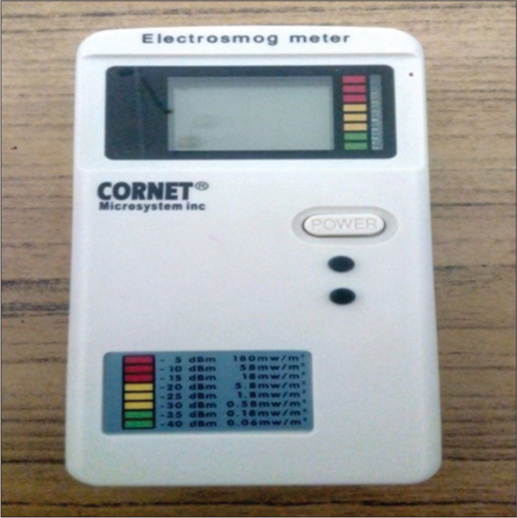
- Radiofrequancy decibel meter.
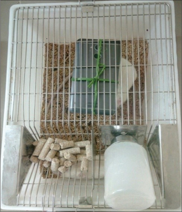
- Rats exposed to Radio Frequency Electro Magnetic Radiations (RFEMR)
Feeding behaviour assessment every day
Food intake was noted in rats. Food was weighed in the beginning and compared at the end. For this experiment, all rats were housed separately.[18]
Measurement of body weight every 10 days
Body weight of the rats was measured using an electronic balance.
Biochemical analysis
At the end of the experimental period, blood samples were collected from all the rats through retroorbital puncture by sterile capillary tubes. Blood was collected in plain tube and it was allowed to clot at room temperature. Serum was separated after centrifugation at 3000 rpm for 10 min. Serum was used for analysis of MDA and serum corticosterone levels.
Serum corticosterone levels
It was estimated by enzyme-linked immunosorbent assay kit method.
MDA
It was estimated by Thiobarbituric acid method using colorimeter.[19]
Data representation and statistical analysis
Data were expressed as mean ± standard deviation. Statistical analysis was carried out using SPSS software. Statistical differences between the groups were evaluated by independent t-test followed by Dunnets comparison test to compare between treated and control groups. Differences yielding P < 0.05 were considered statistically significant.
RESULTS
MDA and serum corticosterone levels were increased in rats exposed to RF-EMR (1.9038 µmoL/L ± 4.034, 0.3836 ± 0.192) compared to controls (1.1465 µmoL/L ± 0.134, 0.0515 ± 0.55). Daily food intake and body weight were decreased in rats exposed to RF-EMR (34.96 g ± 5.61, 265 g ± 23.14) compared to controls (41.04 g ± 5.98, 298.75 g ± 34.095). The results revealed that there was a statistically significant (P < 0.05) increase in the MDA and serum corticosterone levels in RFEMR exposed rats as compared to controls and decrease in the feeding intake in RF-EMR exposed rats compared to controls whereas body weight of the rats was decreased and was not statistically significant.
DISCUSSION
Feeding behaviour is a complex behaviour in our day to day life that is regulated by many mechanisms. However, the exact regulatory mechanisms are not well understood. One of which is found in recent studies is hypothalamus because it is the centre for controlling feeding behaviour. Many factors effects feeding behaviour such as social facilitation, endocrine disorders, seasonal changes and stress. Among them, most common one in our life affecting feeding behaviour is stress from a variety of sources. Recent studies show that RF-EMR emitted from mobile phones is also known to produce stress responses by causing oxidative damage.[1]
According to [Table 1], our study shows that RF-EMR was known to cause statistically significant (P < 0.005) decreased in feeding behaviour and decreased body weight in male SD rats. Similar findings were observed by Carr, he demonstrated that stress inhibits feeding behaviour in all vertebrates. Restraint stresses have been shown to reduce body weight due to decreased intake of food.[20] Favreau-Peigne et al. also demonstrated that chronic stress altered animal welfare but also decreased body weight.[21] Some studies shows that effect of stress on food intake is not dependent on duration but more on the intensity of a stressor. Our study also shows that RF-EMR was known to cause statistically significant (P < 0.002) increase in serum cortisol levels compared to controls.[22] Some studies show that RF-EMR was known to cause oxidative stress. Stress responses can vary in the degree of activation of the hypothalamus-pituitary-adrenal axis and may stimulate either orexigenic (neuropeptide Y [NPY], agouti-related peptide (AgRP)] or anorexigenic (POMC, melanocortin) pathways, in addition to changes in leptin and insulin, thus inhibiting feeding behaviour. Studies show that in terms of appetite regulation, corticotropin-releasing hormone (CRH) is released from the medial parvocellular (mp) paraventricular nucleus of the hypothalamus (PVN) in response to the stressor.[23] In addition to responding to the stressor, adrenocorticotrophine will be released from pituitary and a sequence of events occur to release glucocorticoids. CRH is also released into the arcuate nucleus of the hypothalamus (ARC) to inhibit NPY/AGRP neurons there. This is normally responsible for inhibiting feeding behaviour thus CRH released after stress inhibits appetite.[4]
| Controls (Mean±SD) | RF-EMR (Mean±SD) | P-value | |
|---|---|---|---|
| Feeding behaviour (g) | 41.04±5.98 | 34.96±5.61 | 0.003* |
| Body weight (g) | 298.75±34.095 | 265±23.14 | 0.19 |
| Malondialdehyde (µmoL/L) | 1.1465±0.134 | 1.9038±4.034 | 0.005* |
| Serum corticosterone levels (ng/mL) | 0.0515±0.55 | 0.3836±0.192 | 0.002* |
RF-EMR: Radiofrequency emitted radiations, SD: Standard deviation, *Statistically significant
Other molecules from the CRH family, such as urocortins, also play a role in appetite suppression. Some studies showed that CRH-deficient mice can have normal stress induced suppression of food intake, implicating other CRH-like molecules. Some studies also showed both CRH and urocortins suppress food intake particularly urocortin 1. It is likely urocortins 1, 2 and 3 influence appetite suppression by acting on the CRHR2 receptor in the hypothalamus. Centrally administrated urocortins are also able to suppress ghrelin secretion, potentially preventing ghrelin-induced stimulation of appetite. On the other hand, peripherally administered urocortins act at CRHR2 receptors in the gut to stimulate an increase in circulating ghrelin. In addition to acting on the NPY neurons of the ARC, CRH-induced appetite suppression also involves other regions of the hypothalamus such as the PVN, supra optic nucleus, perifornical and ventromedial hypothalamus as well as brain regions such as lateral septum, para brachial nucleus and the dorsal portion of the anterior bed nucleus of the stria terminalis (BNST). Thus, they demonstrated that CRH injected directly into the dorsal anterior BNST (but not the ventral part or other brain regions such as the central amygdala) significantly reduces food intake.[24]
Target of this research was to scrutinise the effect of RF-EMR on biochemical parameters of oxidative stress associated changes by stress-related determinants, lipid peroxidative activity by MDA levels and serum corticosterone levels. Results of our study indicated that there was significant increase in MDA levels in rats exposed to RF-EMR compared to controls. MDA is marker for neuronal damage and lipid peroxidation, our study proves that RF-EMR may cause oxidative stress which in turn leads to oxidative damage. Oxidative stress and oxidative damages are well established causes of many chronic disorders. [18] Maneesh et al. also found significantly increased MDA levels in testis and epididymis of RF-EMR exposed male rats and finally concluded that mobile RF-EMR induces oxidative stress[22] A study done on rat liver by using antenna radiations suggested that elevated MDA could be due to cytochrome P450-mediated metabolism of the organic hydroperoxide to active alkoxyl radicals that initiated lipid peroxidation (LPO) and led to liver damage. Hence, these metabolic pathways could increase cellular free radicals, which may attack phospholipids, proteins and nucleic acids.[25] The stress of exogenous origins was reported to elevate the cortisol levels in various organisms. [25] Furthermore, we also noticed a significant increase in the serum corticosterone levels of RF-EMR exposed rats compared to the control. In response to the stressor, adrenocorticotrophin is released from the pituitary and a sequence of events is triggered to release the glucocorticoids.[26]
CONCLUSION
Based on findings of our study, we concluded that exposure to mobile RF-EMR leads to increased MDA and serum corticosterone levels causing oxidative stress and decreased feeding behaviour.
Declaration of patient consent
Institutional Review Board (IRB) permission obtained for the study.
Conflicts of interest
There are no conflicts of interest.
Financial support and sponsorship
Nil.
References
- Effect of radio-frequency electromagnetic radiations (RF-EMR) on passive avoidance behaviour and hippocampal morphology in Wistar rats. Ups J Med Sci. 2010;115:91-6.
- [CrossRef] [PubMed] [Google Scholar]
- Alphotochopherol protects against oxidative stress in the fragile knock out mouse: An experimental therapeutic approach for the Fmr1 deficiency. Neuropsychopharmacology. 2009;34:1011-26.
- [CrossRef] [PubMed] [Google Scholar]
- Comprehensive survey of condition-specific reproductive isolation reveals genetic incompatibility in yeast. Nat Commun. 2015;6:7214.
- [CrossRef] [PubMed] [Google Scholar]
- Mobile phone-induced myocardial oxidative stress: Protection by a novel antioxidant agent caffeic acid phenethyl ester. Toxicol Ind Health. 2005;21:223-30.
- [CrossRef] [PubMed] [Google Scholar]
- A bistable Rb-E2F switch underlines the restriction point. Nat Cell Biol. 2008;10:476-82.
- [CrossRef] [PubMed] [Google Scholar]
- A study on the role of antioxidant Vitamin E supplementation on behavioral changes induced by immobilization stress in mice. Indian J Sci. 2012;2:27-30.
- [Google Scholar]
- Effect of repeated corticosterone injections and restraint stress on anxiety and depression-like behavior in male rats. Behav Brain Res. 2005;156:105-14.
- [CrossRef] [PubMed] [Google Scholar]
- Genetic influences on impulsivity, risk taking, stress responsivity and vulnerability to drug abuse and addiction. Nat Neurosci. 2005;8:1450-7.
- [CrossRef] [PubMed] [Google Scholar]
- A role for brain stress systems in addiction. Neuron. 2008;59:11-34.
- [CrossRef] [PubMed] [Google Scholar]
- Reduced serotonin-1A receptor binding in social anxiety disorder. Biol Psychiatry. 2007;61:1081-9.
- [CrossRef] [PubMed] [Google Scholar]
- Brain regulation of appetite and satiety. Endocrinol Metab Clin North Am. 2008;37:811-23.
- [CrossRef] [PubMed] [Google Scholar]
- Assessment of oxidative stress parameters of brain-derived neurotrophic factor heterozygous mice in acute stress model. Iran J Basic Med Sci. 2016;19:388-93.
- [Google Scholar]
- Developmental expression of neuron-specific enolase immunoreactivity and cytochrome oxidase activity in neocortical transplants. Exp Neurol. 1993;124:208-18.
- [CrossRef] [PubMed] [Google Scholar]
- The glycolytic enzyme enolase is present in sperm tail and displays nucleotide-dependent association with microtubules. Eur J Cell Biol. 2000;79:104-11.
- [CrossRef] [PubMed] [Google Scholar]
- Neuron-specific enolase as a biomarker: Biochemical and clinical aspects. Adv Exp Med Biol. 2015;867:125-43.
- [CrossRef] [PubMed] [Google Scholar]
- Neuron specific enolase: A promising therapeutic target in acute spinal cord injury. Metab Brain Dis. 2016;31:487-95.
- [CrossRef] [PubMed] [Google Scholar]
- Sustained high serum malondialdehyde levels are associated with severity and mortality in septic patients. Crit Care. 2013;17:R290.
- [CrossRef] [PubMed] [Google Scholar]
- Oxidative stress, aging, and diseases. Clin Interv Aging. 2018;13:757-72.
- [CrossRef] [PubMed] [Google Scholar]
- Serum MDA levels in myocardial infarction and chronic renal failure. IRCS Med Sci. 1986;14:1110-1.
- [Google Scholar]
- Stress, neuropeptides, and feeding behavior: A comparative perspective. Integr Comp Biol. 2002;42:582-90.
- [CrossRef] [PubMed] [Google Scholar]
- Emotionality modulates the effect of chronic stress on feeding behaviour in birds. PLoS One. 2014;9:87249.
- [CrossRef] [PubMed] [Google Scholar]
- Radio frequency electromagnetic radiation (RF-EMR) from GSM(0.9/1.8GHz) mobile phones induces oxidative stress and reduces sperm motility in rats. Clinics (Sao Paulo). 2009;64:561-5.
- [CrossRef] [PubMed] [Google Scholar]
- Effect of chronic restraint stress on body weight, food intake and hypothalamic gene expressions in mice. Endocrinol Metab (Seoul). 2013;28:288-96.
- [CrossRef] [PubMed] [Google Scholar]
- Eating behavior and stress: A pathway to obesity. Front Psychol. 2014;5:434.
- [CrossRef] [PubMed] [Google Scholar]
- The effects of electromagnetic fields to whole protein, myelin basic protein, neuron specific enolase profiles and nitric oxide levels in rat brains. Ankara Üniv Vet Fak Derg. 2012;59:175-81.
- [CrossRef] [Google Scholar]
- Effects of 900 MHz electromagnetic field on TSH and thyroid hormones in rats. Toxicol Lett. 2005;157:257-62.
- [CrossRef] [PubMed] [Google Scholar]


