
Translate this page into:
Efficacy of Garcinia indica fractions on mast cell degranulation in experimental allergic conditions
*Corresponding author: Md Sadique Hussain, Uttaranchal Institute of Pharmaceutical Sciences, Uttaranchal University, Dehradun, Uttarakhand, India. sadiquehussain007@gmail.com
-
Received: ,
Accepted: ,
How to cite this article: Dayanand L, Vijayakumar S, Veerapur VP, Nagashree KS, Singh SK, Dua K, et al. Efficacy of Garcinia indica fractions on mast cell degranulation in experimental allergic conditions. Indian J Physiol Pharmacol. doi: 10.25259/IJPP_306_2024
Abstract
Objectives:
Allergic reactions are classified into two categories: Early-phase response (EPR) and late-phase response (LPR). EPRs demonstrate a rapid onset of symptoms resulting from histamine release by mast cells (MCs). The introduction of compound 48/80 (C48/80) to rats induces an EPR by facilitating the release of MC contents. Research suggests that inhibiting MC degranulation may hold promise for curing allergic conditions. This article investigates the ability of hydro-alcoholic fractions of Garcinia indica (HAGI) to inhibit MC degranulation. G. indica, a fruit tree from the Clusiaceae family, is known for its use in pharmaceuticals, nutraceuticals, and various industrial applications. Its health benefits are attributed to its high phytochemical content. The aim of this article is to demonstrate the anti-allergic activity of G. indica fruit fractions using both ex vivo and in vivo experimental models.
Materials and Methods:
HAGI was tested for anti-allergic efficacy using a C48/80-generated MC degranulation paradigm in isolated rat mesentery. A dose of 100 μg/mL was administered to a variety of fractions (F2–F8) of the hydro-alcoholic extract.
Results:
MC degranulation in rat mesenteric tissue was markedly reduced at a concentration of 50 mg/kg with a significance of P < 0.01 and at an elevated concentration of 100 mg/kg with a significance of P < 0.001 by the HAGI (F2–F8) in the ex vivo study at 100 μg/mL. The HAGI F6 fraction, when administered at 50 mg/kg and 100 mg/kg in the in vivo study, significantly suppressed the degranulation of mesenteric MC-mediated by C48/80 in rats. The inhibition was significant at P < 0.001 for both concentrations, indicating a robust effect of the fraction at these doses.
Conclusion:
This study validates the traditional use of G. indica fruit fractions in treating allergic diseases, demonstrating significant MC stabilising activity and antiallergic effects. Further, systematic research is required to clarify the precise fundamental process in the MC stabilising functioning of these fractions.
Keywords
Anti-allergic
Allergic diseases
Garcinia indica
Mast cell stabilisation
Histamine

INTRODUCTION
Allergy is a chronic illness characterised by an inappropriate response to a normally harmless substance known as an allergen. Aeroallergens, including dust mites, mould, tree weed and grass pollen, along with food allergens, including eggs, fish, milk, nuts and wheat, are involved in allergies. The term ‘allergy’ refers to a hypersensitivity disorder in which the immune system reacts to specific chemicals. It is a potentially life-threatening and rapid-onset disease reported in all age groups.[1] Allergic reactions occur in response to different environmental proteins, sometimes called allergens. These reactions may be seen as symptoms such as hay fever, asthma, food allergies, swelling of the skin and eye allergies.[2] Allergy conditions are classified into two phases: The first is the activation and sensitisation of T- and B-cells, which, together with the stimulation of mast cells (MCs), is essential for the emergence of allergic reactions, and the second is the immunoglobulin E (IgE)-dependent invasion of eosinophils. Innate lymphoid cells and triggered CD4+ T helper type 2 (Th2) lymphocytes are what drive these processes. These elements are essential to the process of allergic inflammation, which can lead to tissue damage and serious allergy illnesses.[3] There are several mediators, different cell types, and pathways engaged in the processes linking the allergy inflammatory signallings. While interleukin (IL)-5 is necessary for the development and multiplication of eosinophils, the TH2 lymphocyte modulates the signalling, which results in the release of IL-4 and plays a key part in the generation of IgE antibodies. IgE interacts with the high-affinity IgE receptor (FcRI) on macrophages and basophils to affect the allergic inflammatory response. MCs participate in allergic inflammation due to their potent collection of inflammatory facilitators, which include prostaglandin D2, histamine, tumour necrosis factor -α, leukotriene C4, heptin and IL-4. Tryptase, IL-5, chymase, thromboxane A2, platelet-activating factor and IL-6 are a few of the drugs given. Researchers are actively looking at IgE and IL-4 to develop incredibly effective, cutting-edge treatments for allergy problems.[4] Heredity, sex, race and age all increase the risk of allergies, with heredity being the most significant factor. Passive smoking and pollution may function as adjuvants to allergen exposure. Host and environmental factors are two broad groups of allergy risk factors.[5] Allergies are becoming increasingly complex and severe worldwide, a trend driven by multiple factors such as environmental pollution, changes in diet, urbanisation and heightened exposure to allergens. These factors, combined with genetic susceptibility, have led to a rise in both the incidence and severity of allergic diseases, complicating their management and treatment.[6] Children are the ones who suffer the most from allergic illnesses. Anaphylaxis, which can be fatal; food allergies, some types of asthma, conjunctivitis, rhinitis, urticaria, angioedema, eczema and eosinophilic illnesses, among others, are some examples of allergic diseases. While food allergies impact 200–250 million individuals worldwide, asthma affects 300 million people globally. High-, low- and middle-income countries’ allergy rates are rising. As per the World Health Organisation, 300 million people worldwide are thought to have asthma presently; forecasts show that this number will climb to 400 million by 2025.[7] Over 150 million individuals in Europe suffered from chronic allergic illnesses in 2014, and it is anticipated that by 2025, more than half of the population will be impacted by allergy diseases.[8] Globally, 10–30% of people were suffering from these types of diseases and in India, this was around 20–30% of the population; studies in India also report that allergic rhinitis and asthmatic disorders among children range between 20% and 30%.[9,10] Omalizumab, a monoclonal antibody similar to IgE, attaches to unbound IgE, stopping the stimulation of cells such as MCs, basophils and dendritic cells and reducing the expression of FcRI. Severe eosinophilic asthma may now be managed with monoclonal anti-IL-5 antibodies.[11] Vaccinations have had a substantial influence on worldwide health. While vaccinations usually attempt to create or enhance immune responses, current research has shown that adjuvants may also regulate existing immunity or even inhibit harmful immunological responses.[12] At present, antiallergic drugs used in treatment include antihistamines, MC stabilisers, immune suppressors and corticosteroids, which also work as anti-inflammatory agents by targeting several cytokines. These medications have several adverse effects. Often, the first-line therapy of choice for allergic rhinitis is a histamine H1-receptor antagonist. In addition to blocking histamine, numerous second-generation antihistamines have demonstrated suppressive impacts on additional chemical drivers of inflammation, including kinin, leukotrienes and prostaglandins. While second-generation antihistamines are generally well-tolerated and associated with fewer central nervous system effects, the application of corticosteroids in treating severe allergic conditions introduces additional risks. Prolonged use of glucocorticoids is linked with an elevated hazard of several psychological conditions, including anxiety, depression, mood swings and, in rare cases, glucocorticoid-induced psychosis. The dermatological changes and increased cardiovascular risks associated with long-term corticosteroid use, along with mental health concerns, make such therapies less favourable. Therefore, there is a need for new strategies, such as the use of Garcinia indica fractions, to manage allergic conditions more safely and effectively.[13] These actions are less reasonable, leading to the need for new strategies to treat disorders. Hence, the objective of the ongoing study is to establish therapies that can inhibit, delay or counteract allergic reactions. Medicinal plants have been employed in healing since ancient times. Therefore, owning to its extensive history in traditional healing, proven safety record and widespread availability, herbal remedies continue to be a popular choice for therapeutic applications for prevalent diseases throughout India.[14] At present, various plant-derived extracts, including phenolic compounds and flavonoids, exhibit anti-inflammatory functions by modulating the expression of different inflammatory drivers. Herbal ayurvedic plants such as Abies pindrowroyle, Achyranthes aspera L., Bistorta amplexicaulis, Curcuma longa L., Elettaria cardamomum, Ferula assa-foetida L., Glycyrrhiza glabra L. and Justicia adhatoda shows their therapeutic effect against allergic reactions.[15] The slender evergreen tree G. indica (Clusiaceae) is unique to India’s west coast. The polysaccharide content of the fruit has been documented. Kokum has historically been utilised in herbal remedies for the treatment of diarrhoea, inflammatory conditions, dermatitis, gastrointestinal issues and rheumatic pain and as a preventive measure against excessive perspiration. Fruits have medicinal properties and can be used as anthelmintics and cardiac tonics. Kokum rind juice is employed for managing diarrhoea, dysentery, piles and colic illnesses. The traditional method of using a concentrated liquid extract made from the outer covering of fruits is commonly employed as a remedy for diabetes. G. indica leaves are utilised for the treatment of ulcerated skin, dyspepsia and hyperplasia.[16] Anthocyanins are present in tannin, along with organic acids and anthocyanins. Kokum and its chemical constituents have been shown to possess antifungal, antibacterial, cardioprotective, anti-ulcerogenic, anticancer, chemopreventive, antioxidant, free radical scavenging and anti-lipidaemic properties in preclinical research.[17] Considering all these above scientific data and strong traditional claims available in the literature, the present study assessed the potential stabilising impact of various fractions on MC of G. indica using different experimental animal models.
MATERIALS AND METHODS
Chemicals
Purchased from Sigma Aldrich in Bangalore, India, the compound 48/80 (C48/80) was kept in a deep freeze until required. Obtained from Yarrow Chemicals, the traditional preventive medicine disodium cromoglycate (DSCG) was preserved under the required storage conditions. All other compounds used in this study, including gallic acid, Folin– Ciocalteu reagent, alcohol, acetone, n-hexane, ethyl acetate and methanol, were of analytical grade.
Plant materials, extraction
The fruit rind of the G. indica was obtained from the local region of Sirsi taluk, Uttara Kannada District, Karnataka. The collected fruit samples were identified and authenticated by Prof Shalini B R, Principal, University of Science, Tumakuru University, Tumakuru, and the specimen voucher (Herbarium) is preserved in the department. The fresh fruit of G. indica was collected, seeds were separated, and shade dried until the rind became brittle. Later, the dried fruit rind was ground into a coarse powder and subjected to Soxhlet extraction, using ethanol and water as a solvent in the ratio of 70:30, respectively.
Preparation of fractions from Hydroalcoholic extract of G. indica (HAGI) extract
Different fractions of HAGI were prepared using Column Chromatography. The extract HAGI was redissolved in n-Hexane, mixed with silica gel (60–20), evaporated to dryness, and ground in a mortar and pestle to obtain a homogenous mixture of extract. Column 4.5 × 60 cm was packed with 125 g of silica gel, and the extract bed (5 g of drug extract + 25 g of silica gel) homogeneous mixture was loaded on top of the column.
Animals
Animal subjects used in scientific research Wistar albino rats weighing between 200 and 250 g, both male and female, were obtained from the vivarium of Sree Siddaganga College of Pharmacy. The rats were kept in regular polypropylene cages and treated in typical laboratory settings, with an ambient temperature of 24 ± 1°C, a relative humidity of 55 ± 5% and a 12-h light/dark cycle. The research protocol received clearance from the Institutional Animal Ethics Committee (IAEC) of Sree Siddaganga College of Pharmacy. The approval number is SSCP/IAEC.Clear/206/2020-21. The experimental animals had a 7-day acclimatisation period before the start of the investigation. Selected animals were marked individually and randomised according to bodyweight distribution. So that means the body weight of each group would not be statistically different from those of the groups. The animals were housed in neat polypropylene cages with bedding made of sterile rice husk.
Determination of total phenolic content (TPC) in various fractions of HAGI
To quantify the TPC in HAGI Fractions, the Folin–Ciocalteu colorimetric method was used. Gallic acid solutions in methanol were prepared at various concentrations (6.25, 12.5, 25, 50, 100 and 200 μg/mL). Three hundred microliters of gallic acid at various concentrations were added to each test tube, resulting in a total volume of 1.5 mL. Next, 300 μL of Folin–Ciocalteu reagent (10%) and 900 μL of 7.5% sodium carbonate were introduced. The resulting azure solution was rapidly stirred and left to incubate at room temperature for 120 min. Next, the measurement of absorbance was conducted at a specific wavelength of 760 nm in comparison to a blank sample. The experiment was performed on three separate occasions. A calibration curve was generated by integrating the mean absorbance values acquired at different doses of gallic acid. Furthermore, fractions containing a concentration of 200 μg/mL were generated and their absorbance was quantified using the identical method as the reference. The quantification of the TPC of the fractions was expressed as milligrams of gallic acid equivalents (GAE)/g of dry extract (mg/g). Repeated measurements were conducted 3 times.[18]
Ex vivo and in vivo mesenteric mast cell degranulation (MCD) in rat mesentery
Dose standardisation of DSCG
To evaluate the MC stabilisation impact of DSCG, the medication was diluted in distilled water to create concentrations of 50 μg/mL, 100 μg/mL and 200 μg/mL. These concentrations were selected based on a review of relevant literature and preliminary studies, which indicated that these levels are effective in demonstrating a range of pharmacological responses. The chosen doses span from lower to higher concentrations to assess the dose-response relationship and to identify the minimum effective dose while ensuring safety and feasibility in the animal models. Following a period of fasting, the rats were humanely killed by cervical dislocation. The abdominal cavity was surgically dissected to reveal the intestine. Small portions of the mesenteries were then separated and placed in a Petri dish filled with Ringer Locke solution. The solution consisted of sodium chloride (154 mm), potassium chloride (5.6 mm), calcium chloride (2.2 mm), sodium bicarbonate (6.0 mm) and dextrose (5.5 mm). The Petri dish was kept at a constant temperature of 37°C and provided with aeration. Subsequently, the mesenteric samples were subjected to treatment with different doses of DSCG to stabilize the MCs. The samples were placed on slides and then moved to separate Petri dishes. They were then exposed to specific treatments for 5 min. After that, C48/80 was added to each dish at a concentration of 5 μg/mL, except for the normal control. The incubation was extended by 10 min at a temperature of 37°C. Following the incubation period, the slides containing mesenteric samples were placed in a solution of 4% formaldehyde and 0.1% toluidine blue for 20–30 min. Subsequently, they were treated with xylene for 10 min and rinsed with acetone for 5 min. The discoloured mesenteric samples were analysed using a microscope at a magnification of × 45. A minimum of 100 MCs were tallied, and the proportion of undamaged MCs was calculated. The proportion of protection against MC degranulation was also computed. [19-21]
Standardisation of fractions dose
The Mesenteric Pans were affixed onto slides and then treated with several concentrations (50, 100 and 200 μg/mL) of 100% ethyl acetate HAGI fraction (F3) for 5 min. Subsequently, C48/80 was applied to each Petri dish, excluding the normal control, at a concentration of 5 μg/mL. The dishes were then incubated at 37°C for 10 min. The staining process, counting procedure, and formula are outlined in section 3.1 as follows.
Screening of protective effect of all fractions (F2-F8) of HAGI
The Mesenteric Pans were mounted on slides incubated in different fractions of HAGI (F2-F8) for 5 min. Later, C48/80 (5 μg/mL) was added to every petri dish except normal control and again incubated for 10 min at 37°C. The staining process, counting procedure, and formula are outlined in section 3.1 as follows.
In vivo compound 48/80-mediated MCD on rat mesentery
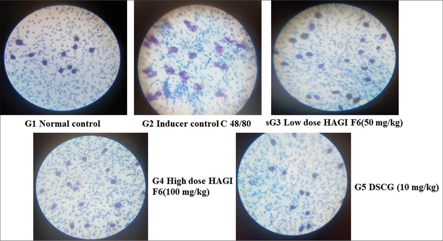
A total of 30 Wistar albino rats, weighing between 180 and 200 g, were placed into five groups, with each group consisting of six animals (n = 6). Group I was given normal saline orally at a dose of 1 mL/kg, whereas Group II, which served as the control for inducing a response, received C48/80 subcutaneously at a dose of 1 mg/kg. On the initial day, rats in Groups III, IV and V were exposed to C48/80 (1 mg/kg, administered subcutaneously) to induce sensitivity. Group III and Group IV were administered the HAGI F6 Fraction, which contained 75% methanol, at low (50 mg/kg, orally) and high doses (100 mg/kg, orally), respectively. Group V received DSCG (10 mg/kg, intraperitoneally) as a benchmark. After 7 days, precisely 2 h following the final dose, the rats were euthanised, and their intestinal mesenteries were gathered to evaluate the stabilisation of MC. The mesenteries, together with small sections of the intestine, were placed on a Petri dish filled with Ringer Locke’s solution at a temperature of 37°C. They were then moved to a slide and stretched using needles. The intestinal tissue was extracted, and the mesenteric samples were exposed to a 5 μg/mL solution of C48/80 for 10 min. Subsequently, they were treated with a solution containing 0.1% toluidine blue in a 4% aqueous formalin solution. The cells that had been labelled were submerged in xylene for a duration of 5–10 min, washed 2–3 times with acetone and examined under a microscope at a magnification of 45× to assess the stabilisation of MC. A total of 100 MCs were observed and quantified across different visual fields. The quantity of cells that were intact and those that had undergone degranulation were documented to determine the percentage of protection.[22]
%protection=(1-[T/C]) × 100
Where T = no. of degranulated cells of the test.
C = no. of the degranulated cell of inducer control
Statistical analysis
A one-way analysis of variance (ANOVA) was conducted using GraphPad Prism 5.1 software for statistical analysis, followed by Tukey’s multiple comparison test as a post hoc study. A statistically meaningful value was defined as P < 0.05.
RESULTS
TPC of Hydroalcoholic extract of G. indica fractions
The TPC of F2 found to be 389.5 ± 64.05 mg GAE/g is the highest TPC fraction and other fractions are F3 = (112.65 ± 1.53), F4 = (44.76 ± 0.58), F5 = (54.17 ± 1.91), F6 = (60.28 ± 2.12), F7 = (31.22 ± 0.22) and F8 = (51.88 ± 1.95) mg GAE/g, respectively. F3 fractions were selected for ex vivo study [Table 1].
Dose standardisation DSCG for Ex vivo study
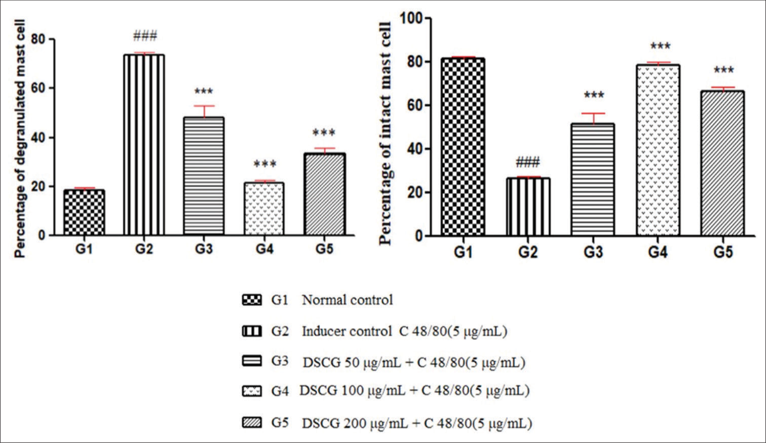
Ex vivo administration of C48/80 (5 μg/mL) produced degranulation of MCs in the mesentery of rats. The percentage of degranulated MCs was substantially increased (P < 0.001) in the C48/80 alone group (73.62 ± 1.10) compared with the normal. Treatment with DSCG (50, 100, 200 μg/mL) exhibited a substantial decrease (P < 0.001) in percentage of degranulated cells (48.22 ± 4.76, 21.3 ± 1.45 and 33.6 ± 2.11, respectively) compared to C48/80 alone group [Table 2 and Figure 1]. Further, the percentage of intact cells substantially increased (P < 0.001) with all the doses of DSCG but 100 μg/mL DSCG exhibited decrease (P < 0.001) in degranulated cells (21.3 ± 1.45) in contrast to two doses (50, 200 μg/mL).
Ex vivo Standardisation of fractions dose
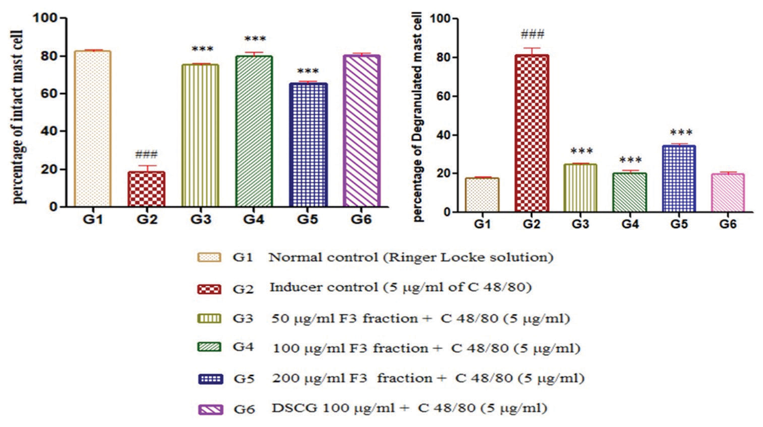
Ex vivo administration of C48/80 (5 μg/mL) produced degranulation of MCs in the mesentery of rats. The percentage of degranulated MCs was increased (P < 0.001) in the C48/80 alone group (81.34 ± 3.73) compared with the normal. Management with F3 fraction (50, 100 and 200 μg/mL) exhibited a significant decrease (P < 0.001) in the percentage of degranulated cells (24.82 ± 0.94, 19.9 ± 1.84 and 34.47 ± 1.04 respectively) compared to C48/80 alone group [Table 3]. MC stabilising activity was shown by all three doses of F3 fractions. It was evident from [Table 3 and Figure 2] that the percentage of intact cells was increased (P < 0.001) with all the doses of 100% ethyl acetate doses of 100 μg/mL HAGI fractions. The F3 fraction demonstrated a significant reduction in degranulated cells, with a mean count of 19.9 ± 1.84, compared to the effects observed with the two doses (50 and 200 μg/mL) of the F3 fraction, which also showed significant effects but were less pronounced (P < 0.001). This indicates that the F3 fraction at the tested concentration was more effective in reducing MC degranulation.
Protective impact of different fractions of HAGI on mesenteric MCD in ex vivo study
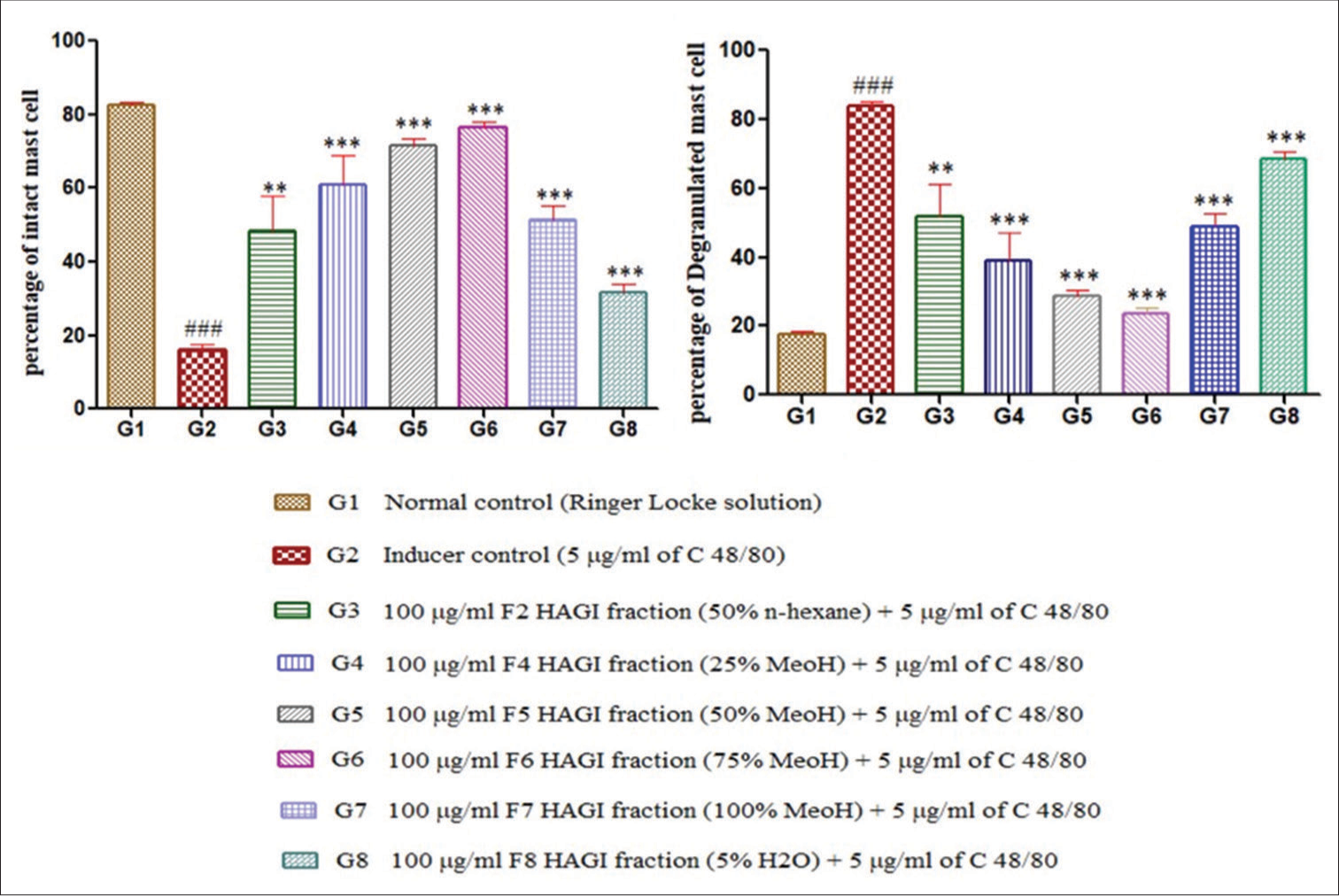
The C48/80 (5 μg/mL) produced degranulation of MCs in the mesentery of rats. The percentage of degranulated MCs was significantly increased (P < 0.001) in the C48/80 alone group (83.83 ± 1.39) compared with the normal group. Challenged with 100 μg/mL for F2 to F8 fractions exhibited decrease (P < 0.001) and (P < 0.01) in percentage of degranulated cells (F2 =51.69 ± 9.69, F4 =39.13 ± 8.10, F5 =28.50 ± 1.76, F6 =23.60 ± 1.74, F7 = 48.62 ± 3.85 and F8 = 68.46 ± 2.26.) compared to C48/80 alone group [Table 4 and Figure 3]. MC stabilising activity was shown by all HAGI fractions with doses of 100 μg/mL. It was evident from [Table 4] that the percentage of intact cells was increased (P < 0.01; P < 0.001) with all the fractions with the doses of 100 μg/mL HAGI fractions. The F6 fraction at a dose of 100 μg/mL leads to significant decrease in degranulated cells, with a mean count of 23.60 ± 1.74, compared to the other HAGI fractions. This reduction was statistically significant (P < 0.001), highlighting the superior efficacy of the F6 fraction in reducing MC among the fractions tested.
In vivo compound 48/80 induced MCD in rat mesentery
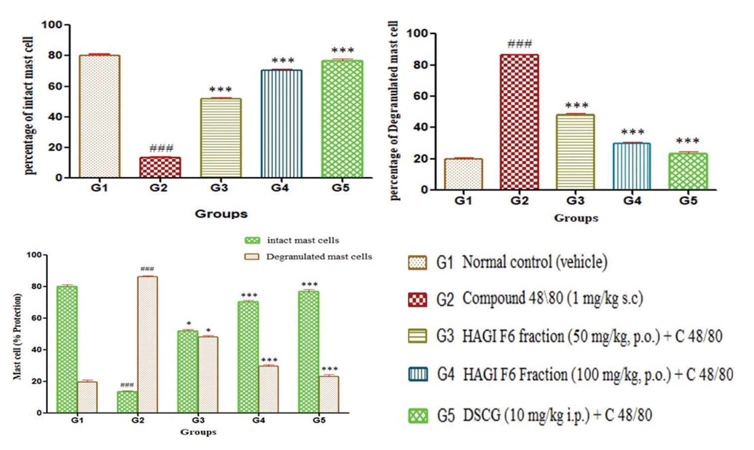
Administering C48/80 subcutaneously at a dosage of 1 mg/kg resulted in the degranulation of mesenchymal cells in the rats’ mesentery. Significantly higher percentages of degranulated MCs (86.45 ± 0.61) were seen in the C48/80 alone group compared to the normal group (P < 0.001). Dosing the F6 fraction of HAGI at doses of 50 and 100 mg/kg resulted in a significant reduction (P < 0.001) in the percentage of degranulated cells (48.09 ± 0.95 and 29.69 ± 1.00, respectively) compared to the C48/80 alone group [Table 5 and Figure 4]. Conventional DSCG also demonstrates a substantial (P < 0.001) reduction in degranulated cells (23.22 ± 1.28) compared to the C48/80 alone group [Figure 5].
| Fractions | Total phenolic content (mg GAE/g) fraction |
|---|---|
| F2: n-hexane: Ethyl acetate (50:50%) | 389.5±64.05 |
| F3: Ethyl acetate (100%) | 112.65±1.53 |
| F4: Methanol: Ethyl acetate (25:75%) | 44.76±0.58 |
| F5: Methanol: Ethyl acetate (50:50%) | 54.17±1.91 |
| F6: Methanol: Ethyl acetate (75:25%) | 60.28±2.12 |
| F7: Methanol (100%) | 31.22±0.22 |
| F8: Water: Methanol (5:95%) | 51.88±1.95 |
GAE: Gallic acid equivalents
| Treatment | Intact mast cells (%) | Degranulated mast cells (%) | Percentage Protection (%) |
|---|---|---|---|
| Normal control (Ringer Locke solution) | 81.52±1.11 | 18.47±1.11 | 80 |
| Inducer control (5 μg/mL of C 48/80) | 26.53±1.06### | 73.62±1.10### | ---- |
| DSCG 50 μg/mL+C 48/80 (5 μg/mL) | 51.78±4.76*** | 48.22±4.76*** | 36 |
| DSCG 100 μg/mL+C 48/80 (5 μg/mL) | 78.64±1.45*** | 21.3±1.45*** | 72 |
| DSCG 200 μg/mL+C 48/80 (5 μg/mL) | 66.4±2.11*** | 33.6±2.11*** | 56 |
DSCG: Disodium cromoglycate, each value represents the mean±standard error of the mean (SEM) (N=5 mesenteric panes). ###P<0.001 compared to normal control; ***P<0.001 compared to inducer Compound 48/80 group. Statistical evaluation was done by one-way ANOVA followed by Tukey’s post hoctest
| Treatment | Intact mast cells (%) | Degranulated mast cells (%) | Percentage Protection (%) |
|---|---|---|---|
| Normal control (Ringer Locke solution) | 82.4±1.03 | 17.58±1.04 | 78 |
| Inducer control (5 μg/mL of C 48/80) | 18.62±3.74### | 81.34±3.73### | ---- |
| 50 μg/mL F3 fraction+C 48/80 (5 μg/ml) | 75.17±0.94*** | 24.82±0.94*** | 69.5 |
| 100 μg/mL F3 fraction+C 48/80 (5 μg/mL) | 80.01±1.84*** | 19.9±1.84*** | 76.8 |
| 200 μg/mL F3 fraction+C 48/80 (5 μg/mL) | 66.52±1.37*** | 34.47±1.38*** | 57.3 |
| DSCG (100 μg/mL)+C 48/80 (5 μg/mL) | 80.23±1.37 | 19.76±1.37 | ---- |
MCD: Mast cell degranulation, DSCG: Disodium cromoglycate, each value represents the mean±standard error of the mean (SEM) (N=5 mesenteric panes). ###P<0.001 compared to Normal control; ***P<0.001 compared to inducer Compound 48/80 group. Statistical evaluation was done by one-way ANOVA followed by Tukey’s post hoctest
| Treatment | Intact mast cells (%) | Degranulated mast cells (%) | Percentage Protection (%) |
|---|---|---|---|
| Normal control (Ringer Locke solution) | 82.40±1.03 | 17.60±1.03 | 78.5 |
| Inducer control (5μg/mL of C.48/80) | 16.15±1.39### | 83.83±1.39### | --- |
| 100 μg/mL F2 HAGI fraction+C 48/80 (5 μg/mL) | 48.31±9.69** | 51.69±9.69** | 38.9 |
| 100 μg/mL F4 HAGI fraction+C 48/80 (5 μg/mL) | 60.87±8.10*** | 39.13±8.10*** | 53.6 |
| 100 μg/mL F5 HAGI fraction+C 48/80 (5 μg/mL) | 71.51±1.75*** | 28.50±1.38*** | 64.2 |
| 100 μg/mL F6 HAGI fraction+C 48/80 (5 μg/mL) | 76.40±1.74*** | 23.60±1.37*** | 72.6 |
| 100 μg/mL F7 HAGI fraction+C 48/80 (5 μg/mL) | 51.38±3.85*** | 48.62±3.85*** | 44 |
| 100 μg/mL F8 HAGI fraction+C 48/80 (5 μg/mL) | 31.54±2.26 | 68.46±2.26 | 19 |
MCD: Mast cell degranulation, HAGI: Hydroalcoholic fractions of Garcinia indica, each value represents the mean±standard error of the mean (SEM) (N=5 mesenteric panes). ###P<0.001 compared to Normal control; ***P<0.001 and **P<0.01compared to inducer Compound 48/80 group. Statistical evaluation was done by one-way ANOVA followed by Tukey’s post hoctest
| Treatment | Intact mast cells (%) | Degranulated Mast cells (%) | Percentage protection (%) |
|---|---|---|---|
| Normal control (vehicle) | 80.18±1.03 | 19.88±1.13 | 78 |
| Compound 48\80 (1 mg/kg s.c) | 13.55±0.61### | 86.45±0.61### | ----- |
| HAGI F6 Fraction (50 mg/kg, p.o.)+C-48/80 (1 mg/kg s.c.) | 51.91±0.95*** | 48.09±0.95*** | 52 |
| HAGI F6 Fraction (100 mg/kg, p.o.)+C-48/80 (1 mg/kg s.c.) | 70.31±1.00*** | 29.86±0.67*** | 73 |
| Disodium chromoglycate (10 mg/kg i.p.)+C-48/80 (1 mg/kg s.c.) | 76.78±1.25*** | 23.22±1.28*** | 82 |
MCD: Mast cell degranulation, HAGI: Hydro-alcoholic fractions of Garcinia indica, each value represent the Mean±standard error of the mean (SEM) (n=6), ###P<0.001 compared to Normal control; ***P<0.001compared to Compound 48/80 group. Statistical evaluation was done by one-way ANOVA followed by Tukey’s post hoctest
- Graph showing mast cell stabilisation activity of different concentrations of disodium cromoglycate on compound 48/80 induced mesenteric mast cell degranulation in ex vivo investigation. DSCG: Disodium cromoglycate. ###: Refers to a comparison between G2 (Inducer control) and G1 (Normal control). It suggests that the differences between G1 and G2 are statistically significant at a high level. This is usually interpreted as p < 0.001, indicating a very significant difference. *: Refers to comparisons between the treated groups (G3, G4, G5) and G2 (Inducer control). The three stars (***) usually indicate a significant difference at p < 0.001, meaning the treatments have a very significant effect compared to the inducer control.
- Graph showing mast cell stabilising effect of different concentrations of F3 hydro-alcoholic fractions of Garcinia indica fraction in compound 48/80-induced mast cell degranulation in rat mesentery in the ex vivo study. DSCG: Disodium cromoglycate. *** indicates p < 0.001, suggesting a highly significant difference compared to the control group (G1 or G2, depending on the comparison being made). indicates p < 0.001 as well but likely for a different comparison, such as the difference between the inducer control (G2) and other groups. This suggests that both *** and ### mark highly significant results, but they might highlight significance in different sets of comparisons: *** likely refers to a comparison between treatment groups (G3-G6) and the inducer control (G2). might specifically indicate the significance of the difference between G1 (normal control) and G2 (inducer control).
- Graph showing mast cell stabilising impact of different fractions of hydro-alcoholic fractions of Garcinia indica in compound 48/80-induced mast cell degranulation in rat mesentery in ex vivo study. HAGI: Hydro-alcoholic fractions of Garcinia indica. *** indicates p < 0.001, representing a highly significant difference compared to the control (G1) or the inducer control (G2), depending on the specific comparison. ** indicates p < 0.01, indicating a significant difference (but slightly less than p < 0.001) in a similar comparison. indicates p < 0.001 for comparisons between the inducer control (G2) and other groups, highlighting a highly significant difference between G2 and other treatments. Thus, *** and ### both denote highly significant results (p < 0.001), but ### seems to be used specifically for comparisons involving the inducer control (G2).
- Graph showing mast cell stabilising effect of different doses of F6 fraction of hydro-alcoholic fractions of Garcinia indica in compound 48/80–induced mast cell degranulation in rat mesentery. HAGI: Hydro-alcoholic fractions of Garcin, DSCG: Disodium cromoglycate. *** denotes p < 0.001, representing a highly significant difference when comparing the treatment groups (G3, G4, G5) to the control groups (likely G1 or G2, depending on the comparison). This indicates that the treatments had a very significant impact on the percentage of intact or degranulated mast cells. also indicates p < 0.001, likely signifying a highly significant difference between the inducer control (G2) and other groups, such as G1 or the treated groups (G3-G5). To summarize: *** shows highly significant differences (p < 0.001) for comparisons between treatment and control groups. highlights a highly significant comparison between the inducer control (G2) and other groups, reflecting the strong difference in the mast cell protection/degranulation effect.
-
In vivo mast cell stabilising effect of F6 fraction of hydro-alcoholic fractions of Garcinia indica and disodium cromoglycate standard drug in Compound 48/80-induced mast cell degranulation in rat mesentery. HAGI: Hydro-alcoholic fractions of Garcin, DSCG: Disodium cromoglycate
DISCUSSION
Inflammatory cells and chemical mediators interact intricately to produce allergic inflammation, which is the result of allergic reactions.[23] The early-phase response (EPR) and the late-phase response (LPR) are the two distinct phases in which these reactions typically manifest. The EPR is distinguished by the degranulation of MCs and the secretion of histamine and other components, including cytokines, whereas the LPR entails the movement of additional inflammatory cells from the blood.[24-26] The sensitisation phase commences when antigen-presenting cells offer allergen fragments to T-cells. This interaction results in the release of IL-4 by T-cells, which drives the generation of allergen-specific IgE antibodies by B lymphocytes. Receptors on tissue MCs are the sites of binding for these IgE antibodies. On subsequent exposure to allergens, these allergens bind to the IgE antibodies, leading to their cross-linking. This cross-linking set off a series of intracellular reactions including tyrosine kinase, phospholipase A2, phospholipase C, protein kinase C and calcium ion influx, finally releasing histamine, leukotrienes and prostaglandins during the EPR.[27]
Traditional anti-allergic medications, such as antihistamines, leukotriene inhibitors, thromboxane A2 inhibitors and corticosteroids, are effective but often associated with adverse side effects. Consequently, there has been a growing interest in natural sources of novel anti-allergic agents. This study focused on evaluating the anti-allergic effects of various fractions of G. indica, particularly through their impact on MC degranulation and inflammation in ex vivo and in vivo models.[28]
Histamine plays a crucial role in allergic reactions, functioning as a vasodilator, constricting smooth muscles and significantly enhancing vascular permeability and the production of respiratory mucus. The attachment of allergens to IgE antibodies on MCs and basophils initiates the release of histamine, resulting in calcium-mediated degranulation.[29,30] C48/80, a polymer used to stimulate MCs independently of IgE, induces MC degranulation by reducing intracellular cAMP levels and increasing intracellular calcium, thereby facilitating histamine release.[31] In our study, we observed that treatment with the HAGI F6 fraction led to a significant reduction in MC degranulation. This reduction was likely due to the suppression of histamine and other inflammatory cytokines in mesenteric cells. The HAGI F6 fraction exhibited a stabilising effect on MC membranes, suggesting its potential as an effective MC stabilizer. The ex vivo studies demonstrated that the HAGI F6 fraction outperformed other fractions in reducing MC degranulation, leading us to select it for further in vivo evaluation. Microscopic analysis indicated that the HAGI F6 fraction delivered at concentration of 50 mg/kg and 100 mg/kg greatly decreased MC degranulation in in vivo experiments. These findings indicate that the HAGI F6 fraction not only stabilises MCs but also demonstrates promising anti-allergic activity compared to standard treatments like DSCG.
Overall, our results suggest that G. indica, particularly its F6 fraction, holds significant promise as a natural anti-allergic agent. Additional investigation is necessary to clarify the specific mechanisms that contribute to its effects and to examine its possible therapeutic uses in the management of allergic conditions.
CONCLUSION
This study evaluates the efficacy of various fractions of G. indica, with a focus on the hydro-alcoholic extract F6 (75% MeOH), in stabilising MCs and modulating allergic responses in investigational animals. The F6 fraction demonstrated a significant ability to stabilize MCs compared to other HAGI fractions. This effect was evident through a marked suppression of mediator release from MCs, highlighting its potential as an effective anti-allergic agent. The stabilising effect of the F6 fraction may be attributed to its potential antihistaminic (H1 antagonist) properties and its ability to prevent histamine release triggered by antigens. This suggests that the F6 fraction impacts the allergic disease course by mitigating the harmful consequences of mediator release. The noted anti-allergic effects of the F6 fraction correspond with the historical applications of G. indica, reinforcing its traditional medicinal claims.
Additional investigation is necessary to clarify the precise mechanisms that contribute to the stabilising role of the G. indica F6 fraction on MCs. This includes detailed investigations into how the fraction interacts with MC membranes and modulates mediator release. The promising results from animal models warrant further clinical studies to assess the safety, efficacy and potential therapeutic applications of the F6 fraction in human subjects. Further investigations should focus on optimising the formulation of the F6 fraction to enhance its efficacy and explore potential synergistic effects with other anti-allergic agents.
Acknowledgements:
The authors are thankful to their institutions for providing support in carrying out the study.
Authors’ contributions
LD: Software, writing – original draft;
SV: Formal analysis, writing – original draft;
VPV: Formal analysis, writing – original draft;
KSN: Supervision, validation and visualisation;
SKS: Software, supervision and visualisation;
KD: Formal analysis, validation;
KP: Software, supervision;
MSH: Software, writing – review and editing;
GG: Formal analysis, supervision, validation.
Ethical approval:
The research/study was approved by the Institutional Review Board at Sree Siddaganga College of Pharmacy, number SSCP/IAEC.Clear/206/2020-21, dated 2020-2021. Declaration of patient consent: Patient’s consent was not required as there are no patients in this study.
Conflicts of interest:
There are no conflicts of interest.
Use of artificial intelligence (AI)-assisted technology for manuscript preparation:
The authors confirm that there was no use of artificial intelligence (AI)-assisted technology for assisting in the writing or editing of the manuscript and no images were manipulated using AI.
Financial support and sponsorship: Nil.
References
- Glycyrrhizic acid inhibits IgE-mediated allergic reactions by controlling immune cells that are involved in allergies. Sci Rep. 2017;7:7222.
- [CrossRef] [PubMed] [Google Scholar]
- Immunological mechanisms of allergen-specific immunotherapy. Nat Rev Immunol. 2006;6:761-71.
- [CrossRef] [PubMed] [Google Scholar]
- Introduction to mechanisms of allergic diseases In: Middleton's allergy essentials. Netherlands: Elsevier; 2017. p. :1-27.
- [CrossRef] [Google Scholar]
- Molecular and cellular mechanisms of allergic disease. J Allergy Clin Immunol. 2001;108:S65-71.
- [CrossRef] [PubMed] [Google Scholar]
- State of world allergy report 2008: Allergy and chronic respiratory diseases. World Allergy Organ J. 2008;1:S4-17.
- [CrossRef] [Google Scholar]
- Allergic diseases and asthma: A global public health concern and a call to action. World Allergy Organ J. 2014;7:12.
- [CrossRef] [PubMed] [Google Scholar]
- A concept of Webgis pollen allergy mapping In: Proceedings of the 17th International Multidisciplinary Scientific GeoConference surveying geology and mining ecology management. Vol 29. 2017. p. :1141-8.
- [CrossRef] [Google Scholar]
- Davos declaration: Allergy as a global problem. Allergy. 2012;67:141-3.
- [CrossRef] [PubMed] [Google Scholar]
- Allergic rhinitis in India: An overview. Int J Otorhinolaryngol Head Neck Surg. 2017;3:1-6.
- [CrossRef] [Google Scholar]
- Pros and cons of the use of antihistamines in managing allergic rhinitis. J Allergy Clin Immunol. 1999;103:S395-9.
- [CrossRef] [PubMed] [Google Scholar]
- Psychiatric adverse effects of corticosteroids. Mayo Clin Proc. 2006;81:1361-7.
- [CrossRef] [PubMed] [Google Scholar]
- Current status of herbal drug standards in the Indian pharmacopoeia. Phytother Res. 2017;31:1817-23.
- [CrossRef] [PubMed] [Google Scholar]
- Medicinal plants used to treat respiratory tract illness in Kaghan Valley. Himalayan region-Pakistan. SMGE Book; :5.
- [Google Scholar]
- Phytochemicals and bioactivities of Garcinia indica (Thouars) Choisy-A review In: Diversity of Garcinia species in the Western Ghats: Phytochemical perspective. Thiruvananthapuram: Jawaharlal Nehru Tropical Botanic Garden and Research Institute; 2016. p. :142.
- [Google Scholar]
- Phytopharmacopoeial specifications of Garcinia indica fruit rinds. Pharmacogn J. 2012;4:23-8.
- [CrossRef] [Google Scholar]
- Antioxidant and nephroprotection activities of Combretum micranthum: A phytochemical, in-vitro and ex-vivo studies. Heliyon. 2019;5:e01365.
- [CrossRef] [PubMed] [Google Scholar]
- Various screening methods for anti-allergic activity: An overview. Int J Pharm Sci Nanotechnol. 2010;3:906-11.
- [CrossRef] [Google Scholar]
- Comparative study of mast cell stabilizing effect of extract of selected plants with proven h1 antagonist activity. World J Pharm Res. 2015;4:981-93.
- [Google Scholar]
- Quantitative determination of mast cell fragmentation by compound 48/80. Br J Pharmacol Chemother. 1954;9:494-7.
- [CrossRef] [PubMed] [Google Scholar]
- Antihistaminic and mast cell stabilizing activity of Striga orobanchioides. J Ethnopharmacol. 2001;76:197-200.
- [CrossRef] [PubMed] [Google Scholar]
- Mediators of inflammation and the inflammatory process. J Allergy Clin Immunol. 1999;103:S378-81.
- [CrossRef] [PubMed] [Google Scholar]
- The mast cell as a source of cytokines in asthma. Ann NY Acad Sci. 1996;796:272-81.
- [CrossRef] [PubMed] [Google Scholar]
- Antihistaminic activity of aqueous extract of stem bark of Ailanthus excelsa Roxb. Pharmacogn Res. 2011;3:220.
- [CrossRef] [PubMed] [Google Scholar]
- An update on the application of nano phytomedicine as an emerging therapeutic tool for neurodegenerative diseases. Curr Bioact Compounds. 2024;20:78-91.
- [CrossRef] [Google Scholar]
- Lipoic acid suppresses compound 48/80-induced anaphylaxis-like reaction. Anat Cell Biol. 2010;43:317-24.
- [CrossRef] [PubMed] [Google Scholar]
- The mechanisms of compound 48/80-induced superoxide generation mediated by A-kinase in rat peritoneal mast cells. Biochem Mol Med. 1997;61:107-13.
- [CrossRef] [PubMed] [Google Scholar]
- Studies on the novel antiallergic agent HSR-609: Its penetration into the central nervous system in mice and guinea pigs and its selectivity for the histamine H1-receptor. Jpn J Pharmacol. 1997;73:291-8.
- [CrossRef] [Google Scholar]
- From nature to therapy: Luteolin's potential as an immune system modulator in inflammatory disorders. J Biochem Mol Toxicol. 2023;37:e23482.
- [CrossRef] [PubMed] [Google Scholar]
- Mast cells signal their importance in health and disease. J Allergy Clin Immunol. 2018;142:381-93.
- [CrossRef] [PubMed] [Google Scholar]


