
Translate this page into:
Unveiling the therapeutic potential of Olax psittacorum: An approach to explore its safety and efficacy in experimental rats
*Corresponding author: Lopamudra Adhikari, Department of Pharmaceutical Sciences, S’O’A University, Kalinga Nagar, Bhubaneswar, Odisha, India. lopamudraadhikari@soa.ac.in
-
Received: ,
Accepted: ,
How to cite this article: Mansingh PP, Adhikari L, Dhara M. Unveiling the therapeutic potential of Olax psittacorum: An approach to explore its safety and efficacy in experimental rats. Indian J Physiol Pharmacol. doi: 10.25259/IJPP_157_2024
Abstract
Objectives:
A traditional herb, Olax psittacorum (Lam.) Vahl. used in Odisha, India, for the management of different ailments, including aphthae ulcers. O. psittacorum leaf ethanolic extract was used in the present study to quantitatively evaluate the phytoconstituents present in ethanolic extract for its antibacterial, anti-oral-ulcer and free radical-scavenging properties.
Materials and Methods:
The leaves were extracted with ethanol, and the quantitative estimations were assessed by total tannin, flavonoid as well as phenolic contents. The antioxidant efficacy was evaluated using different free radical scavenging assays, and antimicrobial activity was assessed for Gram-positive as well as Gram-negative bacteria. Acute toxicity and sub-chronic toxicity studies were assessed. The anti-oral-ulcer property of the extract was observed using an acetic acid-induced ulceration in vivo model.
Results:
The extract yield was 45.6%, and the total phenolic, flavonoid and tannin content was determined to be 97.6 mg gallic acid equivalent/g, flavonoid content was 754.8 mg quercetin equivalent/g of extract, and the total tannin content was 21.81 mg tannic acid equivalent/g of extract. The extract exhibited free radical scavenging efficacy with a median inhibitory concentration of <50 µg/mL. Acute toxicity studies revealed no death associated with the extract. Subchronic toxicity studies showed no significant changes in biochemical, haematological or histological parameters. However, the extract exhibited good antioxidant, antimicrobial and anti-oral-ulcer activity by inhibiting ulceration by 70% and 83%, with doses of 100 and 200 mg/kg, respectively, on day 5.
Conclusion:
The present work confirms potent antioxidant and antimicrobial, along with anti-oral-ulcer properties and is considered safe for therapeutic purposes, suggesting that it could be an appropriate herbal source in treating associated ailments.
Keywords
Olax psittacorum
Antioxidant
Quantitative analysis
Antimicrobial
Oral ulcer

INTRODUCTION
Aphthae, mouth ulcers, recurrent aphthous stomatitis (RAS), ulcers or canker sores are confined to the mouth and frequently cause inflammation. Oral or Aphthous ulceration, an ailment characterised by damage to intraoral mucosa, is a prevalent disorder with unknown aetiology that may lead to significant discomfort and pain, as well as masticatory function and altered speech in those affected.[1] It is distinguished by the presence of one to numerous rounded, shallow ulcers of varying sizes on the labial and buccal mucosa, the surface of the ventral tongue and the mouth.[2] Lesions heal naturally within 7–14 days without any scarring. They may reoccur occasionally or frequently, but usually in distinct places.[3] Anxiety, food hypersensitivity, stress, transient weakness, trauma, hormonal fluctuations, Vitamin B12 deficiency, mechanical irritations, iron, folate, ferritin and other medications have been associated with the development of RAS.[4] These medications include cancer chemotherapy, barbiturates, aspirin, phenytoin, penicillin, streptomycin and sulphonamides. No single condition has been found to be the exact cause of stomatitis, nor has an exact aetiology been ascertained. Recurrent aphthous ulcer affects about 20% of adult populations in the United States.[5] The extent of the lesions establishes management, but in all cases, the prevalent recommendation regarding treatment is to reduce the pain and duration of ulcers by suppressing the immune response locally and preventing further infection.[6] Patients with severe or frequent RAS are advised to receive systemic immunosuppressive treatment; however, prolonged administration of these drugs may lead to drug resistance, an imbalance in the oral flora and a secondary bacterial or fungal infection.[7] The absence of adverse consequences in natural extracts has proven their viability as a treatment regimen.[8,9] There are still issues with effectiveness despite the numerous therapeutic protocols that have been attempted.[10,11] Secondary metabolites found in natural extracts include flavonoids, polyphenols and lipophilic, water-soluble polysaccharides. These metabolites stimulate the immune system and are related to anti-inflammatory and anti-adherence effects.[12] Natural extracts have been shown in clinical studies to reduce mouth ulcer pain and duration, but there is insufficient evidence based on animal model experiments to evaluate the effectiveness, safety, outcome and adverse effects of potential treatments.[13,14]
Olax psittacorum (Lam.) Vahl, additionally referred to as Fissilia psittacorum or Olax scandens, is an ayurvedic plant used to treat anaemia, diabetes and antipyretics.[15] Its bark has traditionally been employed to treat anaemia, and its leaves and fruits are consumable. Ethnomedicine also encompasses the use of the leaves of the plant as medicine and food, with roasted leaves consumed as a leafy vegetable as well as its tender leaves being chewed as remedies for mouth or oral ulcers.[16] It has been found that leaf aqueous extract of O. psittacorum plant exhibits anti-inflammatory properties. In an animal model, toxicity studies of various aerial parts revealed a safe dose of 200 mg/kg/bw.[17] O. psittacorum plant extracts, with the exception of alkaloids, contain reducing sugar, proteins, amino acids, glycosides, flavonoids, saponins, tannins, steroids and terpenoids that have the potential to exhibit anti-inflammatory, antioxidant as well as anti-ulcer properties.
The purpose of this research was, therefore, to assess the anti-oral-ulcer efficacy of O. psittacorum leaf ethanolic extract (LEE) on acetic acid-induced oral ulcers in vivo, as well as to quantitatively estimate the phytoconstituents present, free radical scavenging activity, along with antimicrobial efficacy. The study investigated a new source of anti-ulcer agents and used statistical analysis to determine the correlation between anti-microbial and antioxidant activity with phytoconstituents present.
MATERIALS AND METHODS
Plant material
Leaves of O. psittacorum were collected from Chandaka Jungle on the outskirts of Bhubaneswar, Odisha. Collected leaves were authenticated by Dr P.C Panda from the Centre of Biotechnology, Bhubaneswar, India, Plant specimen under field No 2402/CBT.
Extraction
Defatted leaves (500 g) were further subjected to soxhelation using ethanol for 72 h to get the leaf ethanolic extract (LEE). The collected extract was filtered and concentrated using a rotary evaporator at 45°C under reduced pressure.[18] Then, reduced concentrates were further lyophilised at −20°C to get dried LEE. The dried extract was stored at 4°C for further use.
Quantitative analysis of the phytoconstituents
Quantitative assessment of the total phenolic content (TPC)
Quantitative estimation of the TPC was assessed using a reagent Folin-Ciocalteu reagent (FCR) followed by gallic acid as the standard.[19,20] 0.5 mL of extract was taken, and then 0.5 mL of gallic acid was added to it; furthermore, 2.5 mL of FCR reagent was mixed, followed by 2 mL of 7.5% sodium carbonate. The yellow-coloured FCR changes to blue in the presence of the phenolic compound. Then, the mixer was kept at room temp for 30 min. Spectrophotometrically absorbance was observed at 760 nm by employing an ultraviolet (UV).[21] All outcomes were presented as mg gallic acid, equivalent (GAE)/g dried leaf extract.[22]
Quantitative assessment of the total tannin content (TTC)
Quantitative assessment of the TTC was estimated using Folin-Denis reagent (FDR) followed by tannic acid as the standard.[20] 1 mL of extract was taken, and 1 mL of tannic acid solution was added to it. In addition, 1 mL of FDR was added, followed by 2 mL of 7.5% sodium carbonate. The previously prepared solutions were properly mixed, and then absorbance was observed spectrophotometrically at 700 nm using UV. All outcomes were presented as mg tannic acid equivalent (TAE)/g dried leaf extract.[22]
Quantitative assessment of the total flavonoid content (TFC)
Quantitative estimation of TFC was estimated using the aluminium chloride process, taking quercetin as the standard.[20] 1 mL of extract was taken, and then 1 mL of quercetin solution was diluted with 4 mL of distilled water.[19] To the prepared solution, 0.3 mL of 10% sodium nitrite solution was mixed, followed by 0.3 mL of the aluminium chloride (10%). Then, the prepared solution was incubated for 6 min at room temp. 2 mL of sodium hydroxide (1%) solution was mixed with it. Then, the absorbance was observed spectrophotometrically at 510 nm using an ultraviolet-visible (UV-VIS) spectrophotometer.[20] All the results were expressed as mg quercetin, equivalent per Gram dry extract.[21]
In vitro antioxidant study
2,2’-diphenyl-1-picrylhydrazyl (DPPH) radical scavenging assay
In the DPPH assay, the 50% inhibition concentration, or IC50, was determined by plotting a graph of percentage inhibition versus extract concentration.[23] The mixture was stirred and permitted to stand at ambient temperature in the darkroom for about 20–30 min before being combined with 4 mL of DPPH (20 g/mL) solution and 1 mL of various extract concentrations (50–300 µg/mL). The reference standard was ascorbic acid.[24] The absorbance was estimated using a UV-VIS Jasco V-630 spectrophotometer at 517 nm. The formula below was used to calculate the percentage of DPPH free radical inhibition.[25]
DPPH free radical percentage yield-(Ac-As/Ac) ×100
Where Ac is the absorbance of DPPH solution without any sample.
As is the absorbance of the DPPH solution with different concentrations of sample.
ABTS (2,2’-azino-bis(3-ethylbenzothiazoline-6-sulfonic acid) radical scavenging assay
For the ABTS (2,2’-azinobis 3-ethylbenzthiozoline-6 sulphonic acid) free radical efficacy study, the stock solution was made by combining 2.45 mM potassium persulphate and 7mM ABTS solution and the combination was then incubated for 12–16 h.[23] To get 0.7 ± 0.05 absorbance at 730 nm, the stock solution of ABTS was diluted with distilled water in an equal ratio (1:1) to the working solution of ABTS. The ABTS solution was then added using the leaf extract at varying concentrations (50–300 µg/mL) and incubated for 10 min at room temperature under darkness. Using an equivalent amount, ascorbic acid was prepared and used as a reference standard.[24] A UV-VIS spectrophotometer was used to measure the prepared solution at 730 nm after 10 min. The formula below was used to calculate the percentage of ABTS free radical inhibition.
ABTS scavenging effect (%) = Ac-As/Ac×100
Where Ac is the absorbance of ABTS.
As is the absorbance of the Sample.
Hydrogen peroxide (H2O2) scavenging assay
In the H2O2 assay, the potential of the leaf ethanolic extract (LEE) to scavenge H2O2 was observed spectrophotometrically at 230 nm, 10 min after the reaction time.[26] A solution of the H2O2 (2 mM) in phosphate-buffered saline (PBS) with a pH of 7.41 was prepared at 20°C. Different concentration of hydrogen-peroxide was measured spectrophotometrically at 230 nm.[27] Then, at 20°C, the H2O2 solution was added to the final concentrations of LEE (50–300 µg/mL). At approximately 10 min, absorbance was observed in the UV-VIS spectrophotometer against a blank solution that contained LEE (50–300 µg/mL) in PBS without H2O2.[27] Using the same amounts, ascorbic acid was prepared and used as a reference standard, and in the UV-VIS spectrophotometer, absorbance was observed at 230 nm after 10 min.[27] The absorbance (at 230 nm) of the reaction mixture was then recorded at 0 min and every 10 min interval up to 40 min. The concentration of the hydrogen peroxide in assay media was measured. For the calculation of the percentage of H2O2 scavenging efficacy, the following equation was used:
H2O2 scavenging effect (%) = Ac-As/Ac × 100
Where Ac is the absorbance of H2O2
As is the absorbance of the sample.
Phosphomolybdate assay (Total Antioxidant Capacity)
The phosphomolybdenum method was used to determine the total antioxidant capacity (TAC) of the samples.[28] 1 mL of reagent solution was shaken with a 0.1 mL aliquot of the sample solution, sodium phosphate [28 mM], sulphuric acid [0.6 M], as well as 4 mM of ammonium molybdate. Test tubes were covered and placed in a 95°C water bath for about 90 min. The absorbance of the reaction mixture was observed at 765 nm after the samples were cooled.[29] Ascorbic acid was taken as standard. Antioxidant potential was calculated as the ascorbic acid equivalent using the formula:
Total capacity of antioxidants (%) = Ac-As/Ac×100
Where Ac is the absorbance of TAC.
As is the absorbance of the sample.
Reducing power assay
The reducing power of LEE was observed using the previously described method (Yen and Duh 1994). The extract was serially diluted (50–300 µg/mL) in (0.2M) of phosphate buffer solution (pH 6.6) containing 1% ferrocyanide. For 20 min, the mixture was allowed to incubate at 50°C. Some amount of this mixture (5 mL) was treated with 10% trichloroacetic acid 2.5 mL and was centrifuged for about 10 min (at 3,000 rpm). The supernatant was then separated, and 2.5 mL of the distilled water containing 1% ferric chloride was added to it (0.5 mL). This mixture’s absorbance was observed at 700 nm.[30,31] In the reducing power assay, the yellow colour of the Fe3+/ferricyanide complex changes to the ferrous form, which is blue or green (depending on the reducing power) and is referred to as Prussian blue in the reducing power assay.[32] Higher absorbance at 700 nm of Fe2+ indicates the reducing power of the sample extract when compared to ascorbic acid.[33] By following Oyaizu’s method, the reductive capability was examined by measuring Fe3+-Fe2+ transformation in the presence of LEE. The existence of reductants, which act as antioxidants by donating a hydrogen atom and fragmenting free radical chains, is usually associated with the reducing power of the extracts.[34]
Antimicrobial study
Microbial strain collection
Microbial strains that included Staphylococcus aureus (ATCC-6538P), Pseudomonas aeruginosa (ATCC-15442), Escherichia coli (ATCC-43888) and Acinetobacter baumannii (ATCC-19606) were collected to evaluate the antibacterial efficacy.
Disc diffusion assay
The antimicrobial activity of leaf ethanolic extract (LEE) against pathogenic bacteria strains was investigated using the Kirby-Bauer disc diffusion method. The antibacterial assay was performed in Sabouraud dextrose agar and Muller-Hinton agar medium. A bacterial lawn was created by spreading culture over agar at 37°C in an incubator for 24 h.[35] Discs dipped in accurate concentrations of the testing samples were evenly spaced on the lawn agar during antibacterial monitoring.
The microbial suspension was dispersed over an agar medium, and sterile discs were placed on Petri plates after being dipped into extract solution concentrations (150 mg/mL).[36] Common antibiotic and ofloxacin (OF) at a concentration of 10 mg/mL were used as the positive control for the present study, and the zone of inhibition attended by the drugs was used as a comparison parameter for the efficacy produced by the leaf extract against each strain of bacteria, respectively. The Petri plates were incubated with various bacterial strains for 24 h at 37°C and 72 h at 30°C. Zones of inhibition created by test samples in the corresponding plates were measured by a measuring scale.
Toxicity studies
Animal experimentation
The adult Wistar albino rats were employed in the experiments weighing 120-190g, which were collected from the animal house of the School of Pharmaceutical Sciences (SPS), Siksha O Anusandhan Deemed to be University, Bhubaneswar. Before performing the laboratory trials, the rats were acclimatised in polypropylene cages and were provided free unlimited access to the water ad libitum or standard rodent diet along with the controlled laboratory conditions like 12:12 h dark/light environment, room temperature 22 ± 2°C along with relative humidity 45 ± 5%. The animals were provided with human care in accordance with the principles of Laboratory Animal Care (1985). The scientific animal experimentations were carried out following ethical guidelines or ideologies approved by the Institutional Animal Ethical Committee of the Siksha ‘O’ Anusandhan University under the research proposal number IAEC/SPS/SOA/118/2022.
Acute toxicity studies
Acute toxicity research was performed on adult Wistar albino rats of both sexes (150–200 g), employing the Organisation for Economic Cooperation and Development (OECD) 420 guideline. The sighting study involved the oral administration of doses in ascending sequence (100, 200, 400 and 2000 mg/kg) using an oral-gavage. All animals were then observed for the first 4 h for any indication of toxicity or alterations in behaviour. The albino rats were then monitored for about 14 days to observe any indications of toxicity.[37] Due to the absence of any toxicity signs in the sighting study, the main study was performed at 200 mg/kg with 5 additional rats. There were no toxicity signs or behavioural changes observed.[38] As a result, the drug was identified as a GHS category 5 drug (OECD guideline 420, 2001).
Subacute toxicity studies
The subacute toxicity research had been performed by following the guidelines of OECD 407. Leaf ethanolic extract (LEE) (50 mg/kg, 100 mg/kg and 200 mg/kg) was administered daily for 28 days. The repeated oral administration of leaf ethanolic extract (LEE) was continued for 28 days, and no death was observed.[37] On the 29th day, the rats were anaesthetised and then euthanised so that a blood sample could be collected through the cardiac puncture for haematological and biochemical analysis. In addition, vital organs, including the heart, lungs, liver, kidney, brain and stomach, were isolated for the evaluation of the overall relative and absolute weight of organs. A histological study of different organs was done to determine any signs of necrosis.[38]
In vivo anti-oral ulcer efficacy studies
Experimental animals
Swiss albino rats (weighing between 120 and 150 g, b.w.) were chosen for the aphthous ulcer or oral ulcer study and stress-free animals were used in the experiment to prevent bias, as stress can cause ulcers to form or delay the healing procedure after drug administration. Animals were acclimatised to the laboratory environment and provided with a nutritious rodent diet.
Animal grouping
The rats were separated into three groups (n = 3): Group 1 – Negative (ulcer-induced; no treatment), Group 2 – Riboflavin, Group 3 – LEE-100 mg/kg/bw as well as Group 4 – LEE-200 mg/kg/bw, respectively.
Induction of ulcer
An ulcer on the upper surface of the tongue was developed after the application of acetic acid (50%) through a soaked round filter paper (with a diameter of 5 mm). All animals were anaesthetised before ulcer induction by chloroform inhalation within the desiccators. Due to its anaesthetic effect, the tongue of the appropriate group of rats was extracted using a tweezer without causing any damage to their mouths.[39]
Drug administration
The initial day after completion of 24 h of induction was designated ‘DAY-1’; besides a single rat from every group was selected to segregate their tongue for the histopathological examination. Group 2 received riboflavin, and Groups 3 and 4 received doses of LEE of 100 as well as 200-mg/kg/bw on ‘Day 1’, respectively, with the exception of Group 1.[39] Continuation of the administration of dose was identified by proper healing surface of the tongue, and specimen of the tongue from the respective group was collected between ‘DAY 3’ and ‘DAY 5’. Since Group 3 demonstrated a properly healed surface of the tongue without any signs of wounding on day 6, compared to the other groups, a dose of LEE was continued until day 5, which is an indication of the curative or healing property in the early phase or shorter duration.[40,41]
Statistical examination
The Statistical Package for the Social Sciences (SPSS, version 20) software was employed in this study. One-way analysis of variance, followed by Least significant Difference (LSD) and Tukey-test in SPSS, was employed to compare significant differences (P ≤ 0.05) between the treatment as well as control groups.
RESULTS
Extraction
The obtained ethanolic extract was a dark-green sticky mass. The percentage (%) yield of ethanolic extract was calculated by employing the percentage yield formula. The overall percentage (%) yield of the extract was found to be 45.66 ± 0.52%.
Quantitative analysis of the phytoconstituents
The quantitative analysis of leaf extract indicates a high flavonoid content in comparison to other metabolites, with TPC 97.6 GAE/g, TTC 21.81 mg TAE/g and flavonoid content 754.8 mg quercetin equivalent/g of extract, respectively, as shown in Table 1.
| Extract | TPC* | TFC* | TTC* |
|---|---|---|---|
| LEE | 97.6 | 754.8 | 21.81 |
In vitro antioxidant study
The antioxidant activity was performed using LEE and ascorbic acid was taken as standard. The IC50 of LEE with the ABTS radical scavenging assay (17.66 µg/mL), DPPH radical scavenging assay (39.86 µg/mL), H2O2 radical scavenging assay (21.58 µg/mL) and phosphomolybdate radical scavenging assay (43.72 µg/mL) is shown Table 2. Higher concentrations of LEE exhibited antioxidant effects similar to those of ascorbic acid at the same concentration and confirmed the existence of antioxidant properties in LEE [Figure 1].
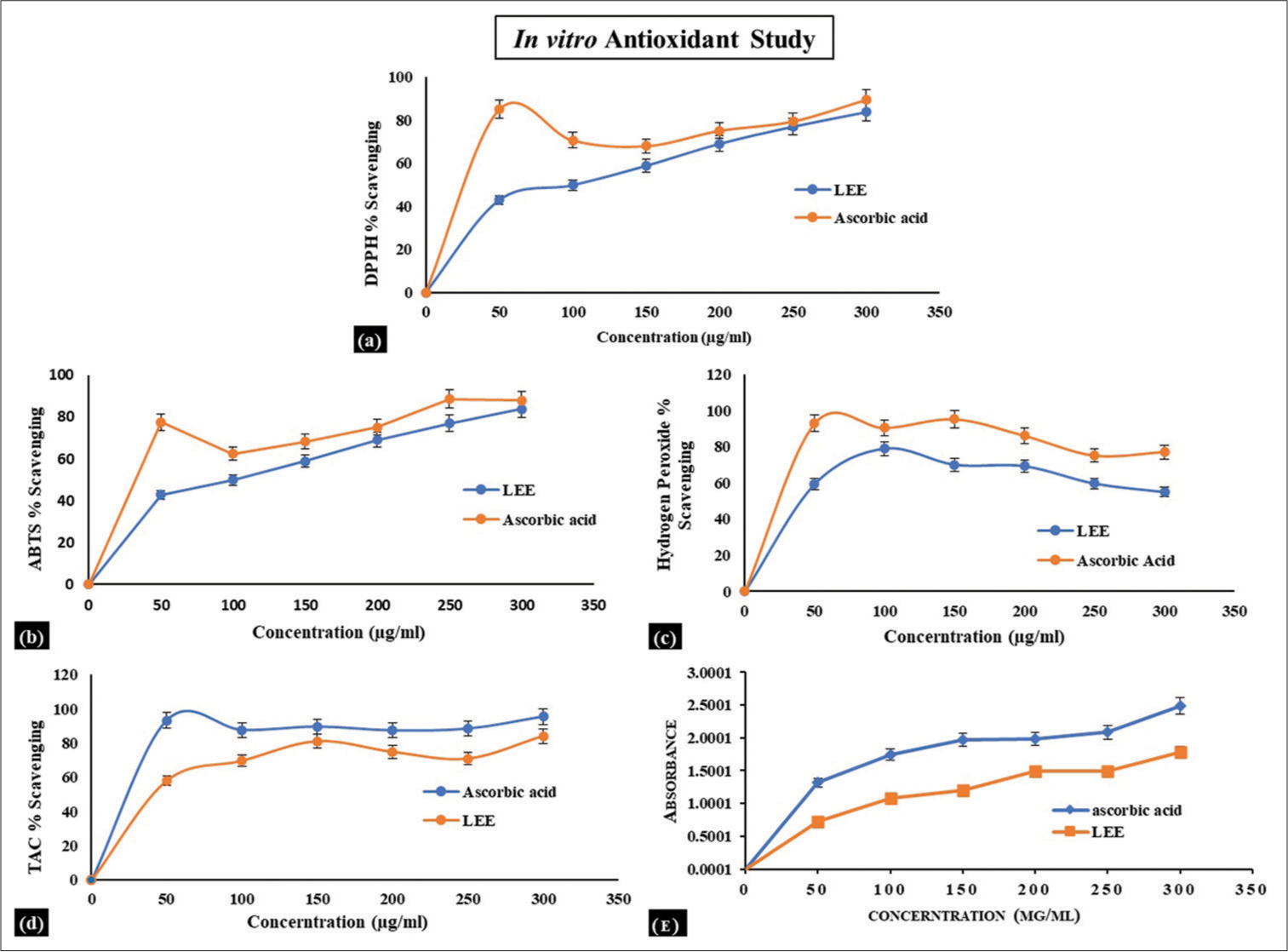
- Observation graph of LEE with the (a) 2,2’-diphenyl-1-picrylhydrazyl radical scavenging activity, (b) ABTS radical scavenging assay, (c) hydrogen peroxide radical scavenging activity, (d) phosphomolybdate radical scavenging activity and (e) reducing power activity.
| Extract | IC50 | |||
|---|---|---|---|---|
| LEE | DPPH scavenging assay | ABTS scavenging assay | H2O2 scavenging assay | Phosphomolybdate assay |
| 39.86 µg/mL | 17.66 µg/mL | 21.58 µg/mL | 43.72 µg/mL | |
DPPH: 2,2’-diphenyl-1-picrylhydrazyl, H2O2: Hydrogen peroxide, ABTS: 2,2’-azino-bis(3-ethylbenzothiazoline-6-sulfonic acid), LEE: Leaf Ethanolic Extract, IC50: Median Inhibitory Concentration
Toxicity studies
Acute toxicity
During the 14 days of observation, no mortality was reported. No cases of morbidity or mortality occurred during the 14 days that followed the dose in the rats, and no toxicity symptoms were noticed. The gross pathological analysis of examined organs revealed no changes. There was no significant variation in relative organ weight [Table 3]. The results of haematology and serum biochemistry of the rats used in the acute toxicity study are presented in Table 4. There was no significant variation observed in the results of LEE groups when compared with the control group. The results of the histopathological study [Figure 2] additionally revealed no abnormalities.
| S. No. | Organs | Control | LEE |
|---|---|---|---|
| Mean of absolute weight | |||
| 1. | Heart | 0.73±0.01 | 0.67±0.01* |
| 2. | Spleen | 0.58±0.01 | 0.51±0.00* |
| 3. | Kidney | 1.43±0.03 | 1.33±0.02* |
| 4. | Adrenal glands | 0.08±0.01 | 0.06±0.01 |
| 5. | Brain | 1.55±0.03 | 1.46±0.02* |
| 6. | Lungs | 1.06±0.02 | 0.96±0.01 |
| Mean of relative weight | |||
| 1. | Heart | 0.48±0.01 | 0.47±0.01 |
| 2. | Spleen | 0.34±0.02 | 0.36±0.03* |
| 3. | Kidney | 0.83±0.01 | 0.82±0.01 |
| 4. | Adrenal glands | 0.06±0.02 | 0.03±0.01 |
| 5. | Brain | 0.93±0.01 | 0.95±0.01* |
| 6. | Lungs | 0.64±0.02 | 0.63±0.03* |
| S. No. | Parameters | Control | LEE |
|---|---|---|---|
| Serum biochemistry | |||
| 1. | Total protein (g/dL) | 6.46±0.01 | 6.47±0.02 |
| 2. | Blood glucose (mg/dL) | 131.06±1.14 | 145.02±2.38* |
| 3. | Serum creatinine (mg/dL) | 0.76±0.02 | 0.68±0.02* |
| 4. | Total bilirubin (mg/dL) | 0.23±0.03 | 0.19±0.01 |
| 5. | Serum cholesterol (mg/dL) | 110.00±1.58 | 122.02±1.48* |
| 6. | Serum triglycerides (mg/dL) | 111.60±1.14 | 142.02±1.92* |
| 7. | Serum calcium (Ca+) (mmol/L) | 11.02±1.30 | 8.06±1.14 |
| 8. | Serum sodium (Na+) (mmol/L) | 137.02±0.83 | 121.80±1.30* |
| 9. | AST (U/L) | 86.06±2.07 | 93.00±1.58 |
| 10. | ALP (U/L) | 73.04±1.14 | 88.00±1.58* |
| 11. | Serum uric acid (mg/dL) | 4.6±1.14 | 3.80±1.30 |
| 12. | ALT (U/L) | 52.40±1.67 | 43.04±1.14* |
| 13. | Serum potassium (K+) (mmol/L) | 4.28±0.08 | 4.62±0.08 |
| 14. | Serum urea (mg/dL) | 45.02±0.83 | 62.02±0.83* |
| 15. | Serum albumin (g/dL) | 4.00±1.22 | 3.08±1.09 |
| 16. | Serum chloride (Cl−) (mmol/L) | 101.04±1.14 | 108.08±1.30* |
| 17. | Serum globulin (g/dL) | 3.32±0.13 | 2.82±0.08 |
| 18. | Blood urea nitrogen (mg/dL) | 8.2±1.48 | 7.08±1.30* |
| Haematological parameters | |||
| 1. | Mean corpuscular haemoglobin (pg/cell) | 17.06±0.89 | 19.00±0.70* |
| 2. | Mean corpuscular volume (fL [µm3]) | 52.02±0.83 | 77.06±0.54* |
| 3. | White blood corpuscles (103/µL) | 4.86±0.05 | 4.58±0.04 |
| 4. | Haemoglobin (g %) | 14.04±0.54 | 09.00±0.70* |
| 5. | Red blood corpuscles (106/µL) | 8.42±0.00 | 6.27±0.01 |
| 6. | Mean corpuscular haemoglobin concentration (%) | 30.24±0.00 | 32.76±0.02* |
| 7. | Packed cell volume (%) | 51.04±0.01 | 48.39±0.03* |
| 8. | Haematocrit (%) | 48.58±0.02 | 48.90±0.07 |
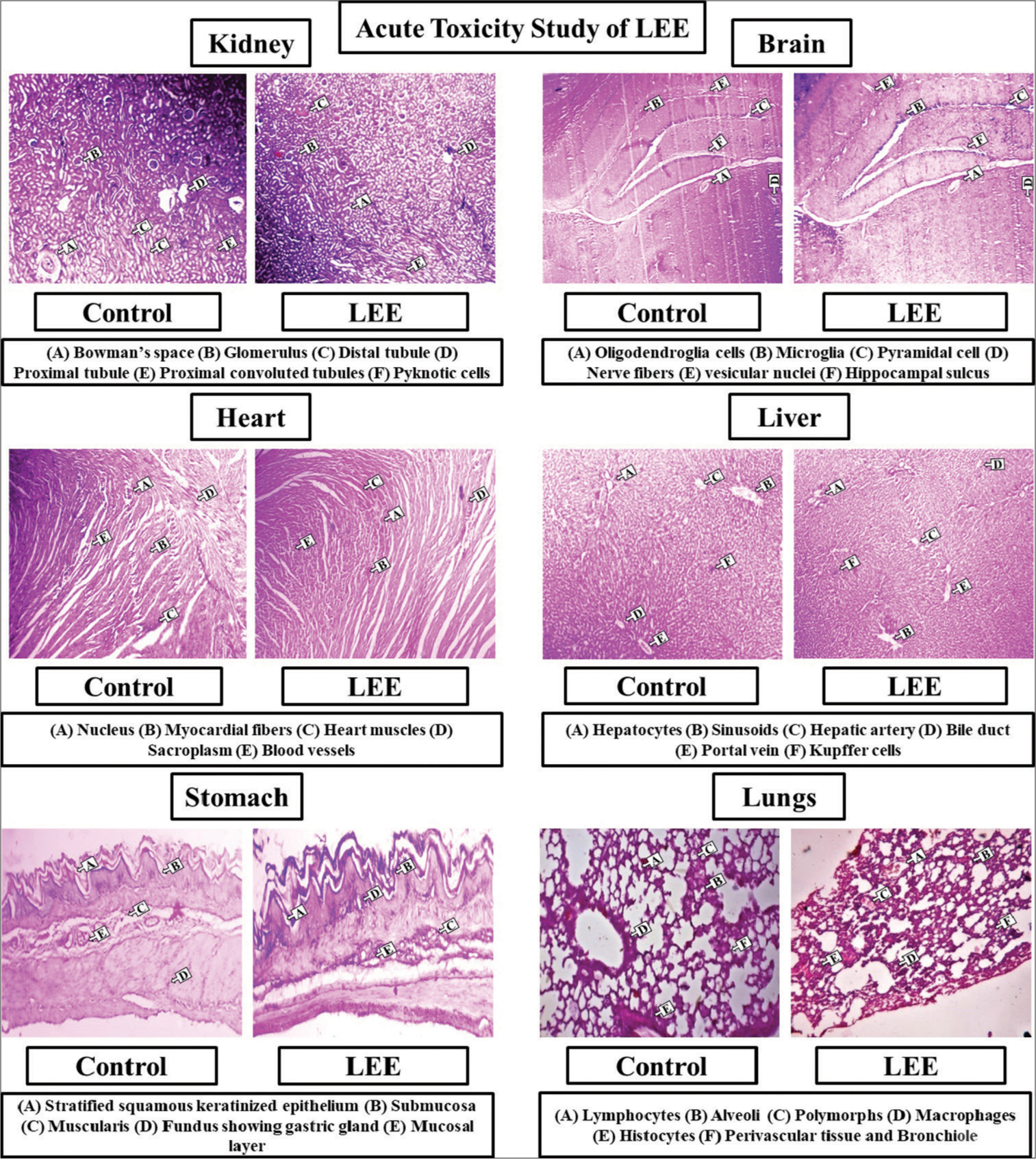
- Histopathological observations of different organs in acute toxicity study of Leaf ethanolic extract (LEE).
In Figure 3, a group of rats that had received LEE treatment had lower body weight than the control group for the first 2–3 days, then it gradually increased. Water consumption for all the rats involved in the study is shown in Figure 3. In addition, the rats who received LEE showed a slight average increase in body weights. Similar to the case with the control group, rats treated with LEE exhibited a steady increase in body weight. The animals given LEE exhibited symptoms resembling diarrhoea that lasted for 2–3 consecutive days. It is probable that the laxative effects of the drug are the cause behind the LEE groups’ lower body weights, lower food intakes and higher stool weights. Figure 3 depicts the food intake of rats throughout the acute toxicity study. With respect to the control group, consistent food consumption was clearly observed. In conclusion to the present study, no gross pathological alterations were observed on the gross necropsy. Table 3 demonstrates the mean absolute weight as well as relative weights (n = 5) of different organs, respectively. The relative weights of organs depicted significant (P ≤ 0.05) differences.
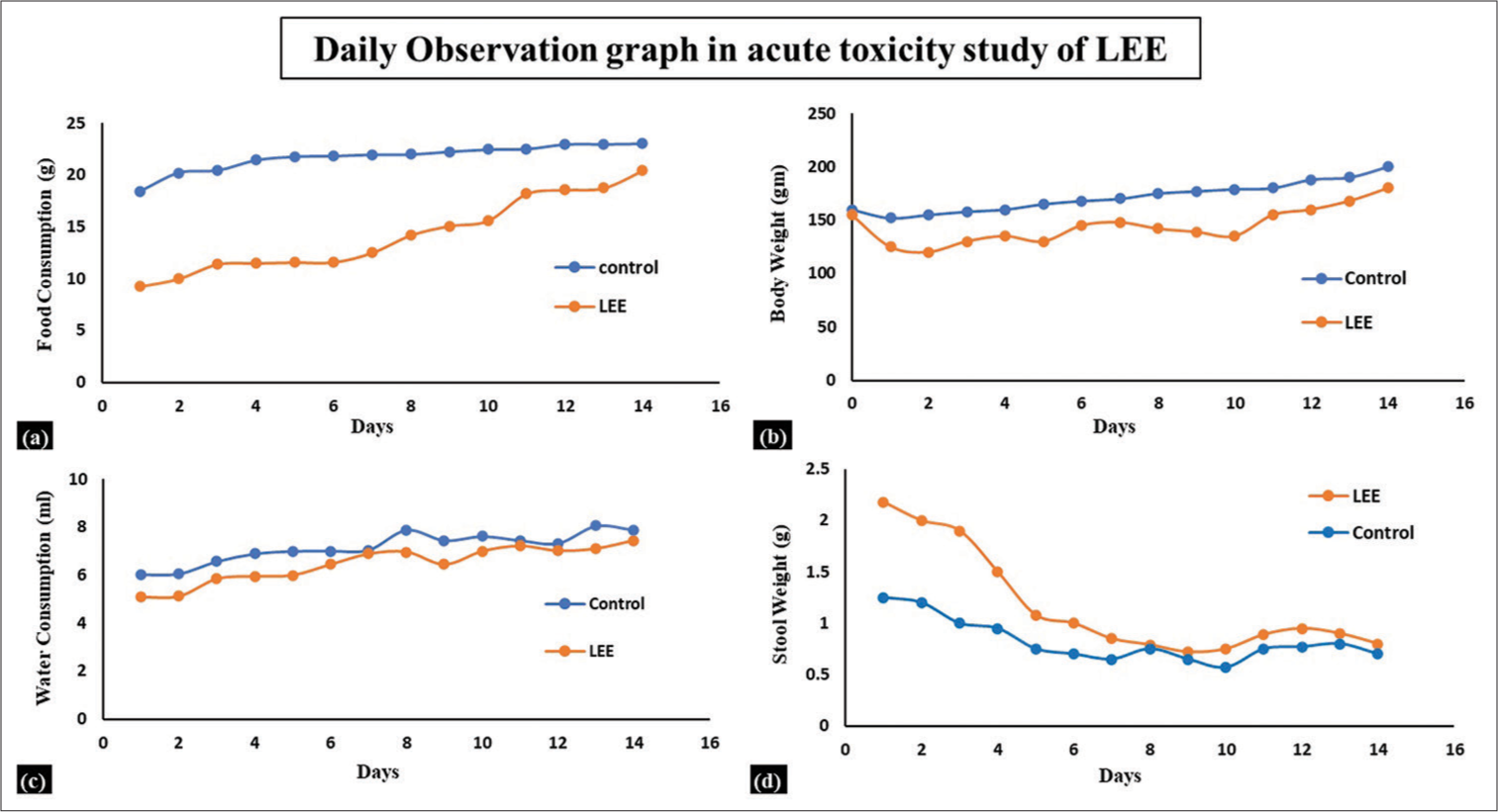
- Daily observation graph in the acute toxicity study of LEE (n = 5). (a) Food consumption, (b) body weight, (c) water consumption and (d) stool weight. LEE: Leaf ethanolic extract.
Sub-chronic toxicity studies
In a sub-chronic toxicity study, LEE was administered to the animals at a dose of 50, 100 as well as 200 mg/kg body weight, respectively. During the study, no group of animals experienced any reduction in body weight. Weekly weight measurements were taken for the treated groups. There was a statistically significant difference (P < 0.05) between the treated groups as compared to the control group in all biochemistry parameters at different dose levels of the extracts [Table 5]. For both the treated and control groups of animals, a haematological investigation revealed the same condition [Table 5]. There was a significant variation observed in the results of LEE groups when compared with the control group. The results of the histopathological study [Figure 4] of different organs additionally revealed no abnormalities. In comparison to the control group, mean relative weight as well as mean absolute weight of animals were observed, where a significant difference was found (P < 0.05) [Table 6].
| S. No. | Parameters | Control | LEE (50 mg/kg) | LEE (100 mg/kg) | LEE (200 mg/kg) |
|---|---|---|---|---|---|
| Serum biochemistry | |||||
| 1. | Total protein (g/dL) | 6.41±0.15 | 6.48±0.19 | 6.59±0.13* | 6.57±0.44 |
| 2. | Blood glucose (mg/dL) | 93.42±3.01 | 90.12±0.54 | 91.76±0.02* | 90.45±2.43 |
| 3. | Serum creatinine (mg/dL) | 0.75±0.04 | 0.71±0.02 | 0.74±0.01 | 0.74±0.04* |
| 4. | Total bilirubin (mg/dL) | 0.46±0.01 | 0.44±0.03* | 0.40±0.01* | 0.43±0.01 |
| 5. | Serum cholesterol (mg/dL) | 101.02±6.58 | 103.12±2.45* | 102.53±1.41 | 104.02±5.32* |
| 6. | Serum triglycerides (mg/dL) | 133.21±2.45 | 132.52±0.02 | 132.06±0.14 | 132.15±3.04* |
| 7. | Serum Calcium (Ca+) (mmol/L) | 10.65±0.12 | 10.23±0.27* | 10.36±0.15* | 10.58±0.29 |
| 8. | Serum sodium (Na+) (mmol/L) | 142.45±12.07 | 143.71±2.45 | 143.06±2.14 | 142.04±2.91* |
| 9. | AST (U/L) | 56.42±6.87 | 56.35±5.21* | 56.40±2.01 | 56.39±3.65 |
| 10. | ALP (U/L) | 82.01±2.75 | 75.46±3.56 | 77.39±2.48* | 78.13±1.29* |
| 11. | Serum uric acid (mg/dL) | 4.75±0.12 | 4.78±0.19 | 4.82±0.45 | 4.89±0.27* |
| 12. | ALT (U/L) | 42.84±6.45 | 39.57±2.89 | 40.63±2.04* | 39.00±2.01 |
| 13. | Serum potassium (K+) (mmol/L) | 4.13±0.45 | 4.12±0.16 | 3.92±0.34 | 3.96±0.12* |
| 14. | Serum urea (mg/dL) | 25.08±1.56 | 23.06±1.96* | 22.47±1.52 | 24.31±1.60 |
| 15. | Serum albumin (g/dL) | 4.97±0.41 | 5.02±0.01* | 5.00±0.09 | 5.01±0.22* |
| 16. | Serum chloride (Cl−) (mmol/L) | 98.01±2.04 | 95.16±2.87 | 94.56±4.12* | 94.78±3.51 |
| 17. | Serum globulin (g/dL) | 3.45±0.04 | 3.42±0.01 | 3.40±4.21* | 3.43±2.01 |
| 18. | Blood urea nitrogen (mg/dL) | 8.95±1.25 | 8.90±0.01 | 8.92±2.03 | 8.90±0.02* |
| Haematological parameters | |||||
| 1. | Mean corpuscular haemoglobin (pg/cell) | 16.54±0.12 | 17.41±1.27 | 17.38±0.55 | 17.36±1.29* |
| 2. | Mean corpuscular volume (fL [µm3]) | 49.85±3.58 | 54.12±3.45* | 53.41±2.84 | 56.58±3.93 |
| 3. | White blood corpuscles (103/µL) | 10.53±0.25 | 10.61±0.33 | 10.69±0.32* | 10.77±0.33* |
| 4. | Red blood corpuscles (106/µL) | 9.75±0.28 | 9.10±0.64 | 9.12±0.23* | 9.08±0.15* |
| 5. | Mean corpuscular haemoglobin concentration (%) | 33.96±2.48 | 33.42±1.09 | 33.28±0.59 | 31.09±0.98* |
| 6. | Packed cell volume (%) | 48.58±2.14 | 49.67±1.65* | 48.23±1.96 | 48.17±1.49 |
| 7. | Haematocrit (%) | 48.59±0.91 | 48.63±1.09 | 48.71±2.45* | 48.91±2.08* |
| 8. | Haemoglobin (gm %) | 16.28±0.84 | 16.29±0.45* | 16.11±0.19 | 16.13±0.64 |
| S. No. | Organs | Control | LEE (50 mg/kg) | LEE (100 mg/kg) | LEE (200 mg/kg) |
|---|---|---|---|---|---|
| Mean of absolute weight | |||||
| 1. | Liver | 6.88±0.01 | 6.40±0.00* | 6.29±0.01 | 6.39±0.04* |
| 2. | Adrenal glands | 0.09±0.00 | 0.04±0.01 | 0.07±0.03* | 0.07±0.01 |
| 3. | Lungs | 1.13±0.01 | 0.95±0.02 | 1.02±0.07 | 0.98±0.02* |
| 4. | Kidney | 1.56±0.07 | 1.50±0.02 | 1.52±0.04 | 1.54±0.03 |
| 5. | Spleen | 0.63±0.02 | 0.56±0.04* | 0.60±0.03 | 0.58±0.04 |
| 6. | Heart | 0.73±0.03 | 0.67±0.02 | 0.70±0.01* | 0.68±0.02 |
| 7. | Brain | 1.78±0.02 | 1.67±0.01 | 1.70±0.00 | 1.69±0.01* |
| Mean of relative weight | |||||
| 1. | Liver | 3.89±0.01 | 3.90±0.00* | 3.93±0.01 | 3.94±0.02* |
| 2. | Adrenal glands | 0.03±0.01 | 0.03±0.00 | 0.03±0.01 | 0.03±0.01 |
| 3. | Lungs | 0.59±0.01 | 0.58±0.00 | 0.57±0.02* | 0.58±0.00 |
| 4. | Kidney | 0.88±0.00 | 0.90±0.01 | 0.91±0.00 | 0.93±0.00* |
| 5. | Spleen | 0.35±0.00 | 0.35±0.01 | 0.35±0.00 | 0.35±0.00 |
| 6. | Heart | 0.40±0.00 | 0.39±0.00* | 0.40±0.01 | 0.40±0.00 |
| 7. | Brain | 0.97±0.02 | 0.98±0.01 | 0.97±0.01 | 0.99±0.00 |
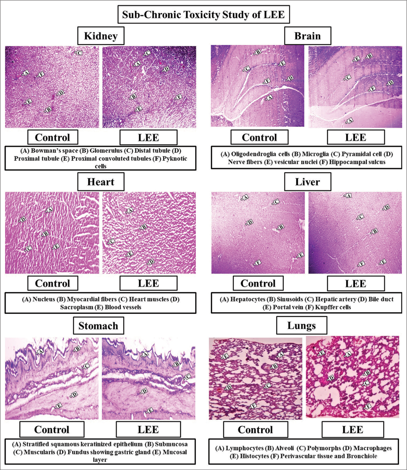
- Histopathological observations of different organs in sub-chronic toxicity study of LEE (Leaf ethanolic extract).
In vivo anti-oral ulcer efficacy studies
The oral ulcer was produced on the tongue of each rat on Day 1 using the approach outlined above [Figure 5]. The LEE 100 as well as 200 mg/kg/bw groups both demonstrated proper healing or cure by Day 5 when compared to the group not receiving treatment or control group, whereas the LEE 200-mg/kg/bw group demonstrated appropriate healing by Day 3 when compared to the LEE 100-mg/kg/bw treated group. On Day 5, LEE 200-mg/kg/bw dose demonstrated proper mucosal or epithelial layer growth as compared to the riboflavin group, indicating appropriate healing of the tongue surface with no indications of injury.
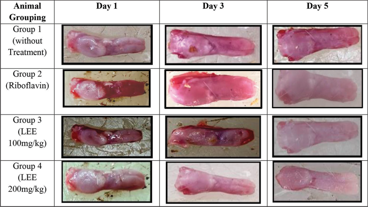
- Induction of ulcer using acetic acid on the tongue of the rats. LEE: Leaf ethanolic extract.
The ulcer index decreased with an increase in LEE dose. There was no significant difference between the ulcerated control rats and the LEE (100 mg/kg) group. In comparison to the ulcerated control (4.82 ± 0.10) and riboflavin (2.03 ± 0.10) groups, the LEE-100 and 200 mg/kg groups significantly decreased the ulcer index by (2.4 ± 0.08) and (1.56 ± 0.04), respectively. In comparison with the rats treated with riboflavin (78%), the ulcer inhibition percentage at LEE-100 mg/kg (70%) and LEE-200 mg/kg (83%) doses was significantly higher [Figure 6]. Hence, the LEE was proven to have good anti-oral-ulcer activity by inhibiting ulceration by Day 5.
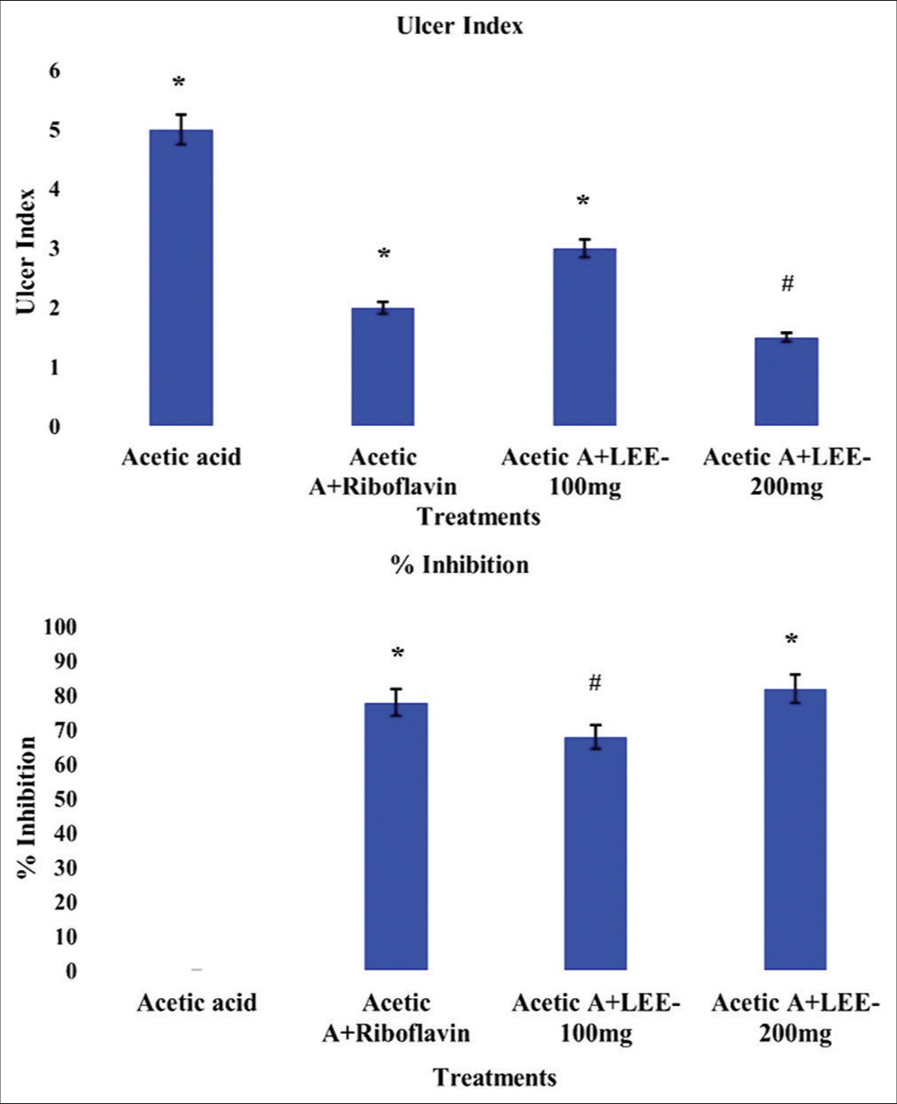
- Effect of Leaf ethanolic extract (LEE) on the ulcer index and effect of LEE on degree of % protection of ulceration. The values were expressed as mean ± standard deviation (n = 6), *P ≤ 0.05 versus respective control group, #P ≤ 0.05 versus respective treatment groups.
The disruption and normal development of tongue cells for the corresponding oral doses are illustrated in the histopathological investigation of the tongue for groups of rats and days [Figure 7].
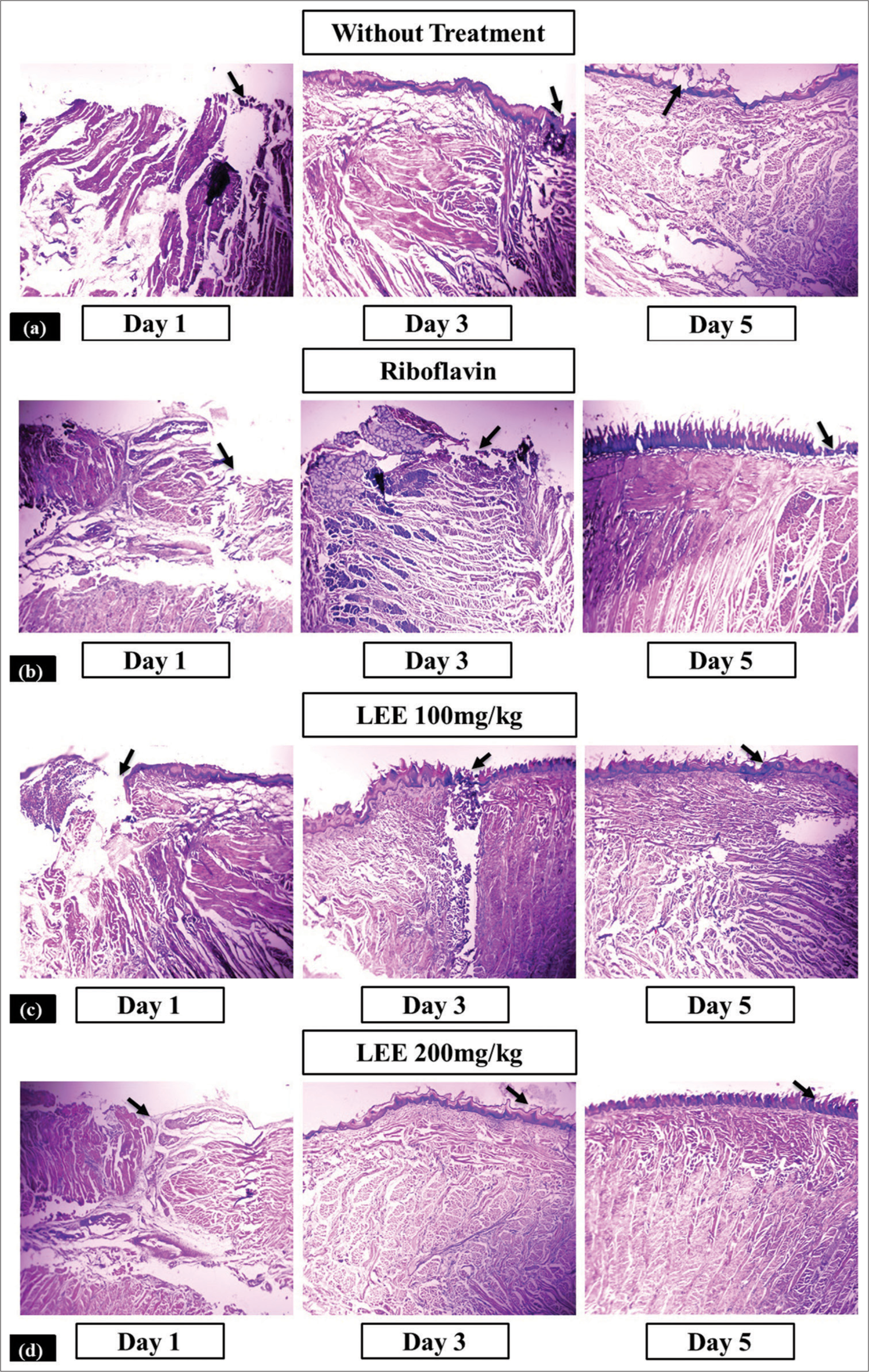
- Histopathological analysis of the isolated tongue samples collected from rats on Day 1, Day 3 and Day 5 to evaluate progress in ulcer healing between different treatment groups, (a) Control (ulcer-induced; no treatment) (b) Acetic acid + Riboflavin, (c) Acetic acid + Olax psittacorum leaf ethanolic extract (LEE-100 mg/kg, p.o.) and (d) Acetic acid + O. psittacorum leaf ethanolic extract (LEE; 200 mg/kg; p.o.). Black arrow in the figures indicates disruption and normal development of epithelial cells of the tongue. LEE: Leaf ethanolic extract.
Antimicrobial study
Using a disc diffusion assay, the leaf ethanolic extract (LEE) was subjected to in vitro antibacterial experiments against Gram-positive and Gram-negative bacterial strains. The findings were compared to those obtained with one common antibiotic OF. A good zone of bacterial inhibition has been seen in the actions of various LEE concentrations.[35] In addition, LEE displayed the best inhibitory action against E. coli and A. baumannii [Figure 8], ranging between 6 mm and 20 mm [Table 7]. Thus, leaf ethanolic extract had generally shown stronger antibacterial activity.
| S. No. | Tested organisms | Zone of inhibition diameter (mm) of LEE at different concentrations | Antibiotic | ||||
|---|---|---|---|---|---|---|---|
| 10 µL (mm) | 20 µL (mm) | 30 µL (mm) | 40 µL (mm) | 50 µL (mm) | OF | ||
| 1. | Escherichia coli | ||||||
| Gram-negative | 6.04±0.01 | 7.02±0.00 | 8.01±0.2 | 10.0±0.26 | 12.04±0.01 | 10.80±0.45 | |
| 2. | Staphylococcus aureus | ||||||
| Gram positive | 4.01±0.10 | 4.06±0.0 | 5.02±0.43 | 6.80±0.51 | 7.01±0.09 | 18.42±0.68 | |
| 3. | Pseudomonas aeruginosa | ||||||
| Gram negative | 5.03±0.71 | 6.04±0.20 | 6.10±0.0 | 7.01±0.0 | 8.26±0.32 | 4.80±0.84 | |
| 4. | Acinetobacter baumannii | ||||||
| Gram negative | 13.01±0.60 | 14.08±0.02 | 16.03±0.06 | 17.01±0.46 | 19.04±0.28 | 0 | |
OF: Ofloxacin, LEE: Leaf ethanolic extract
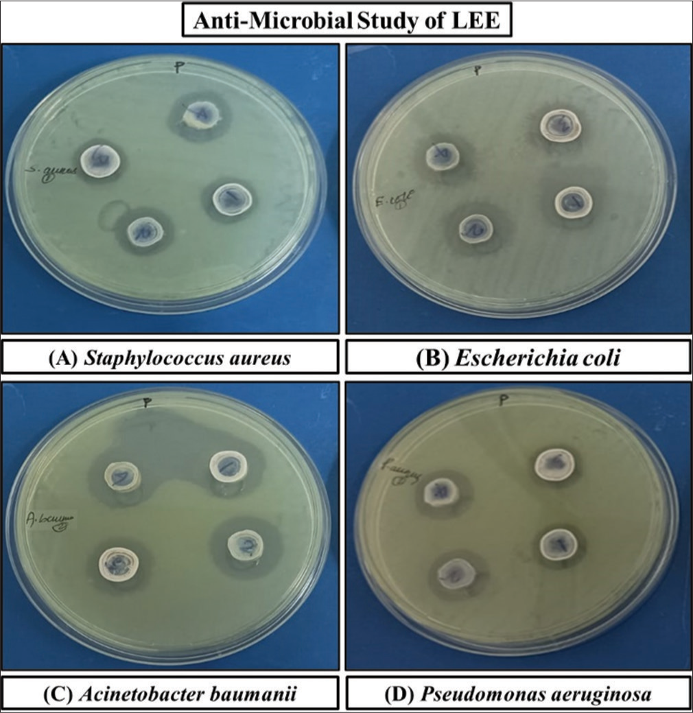
- Antibacterial activities of Leaf ethanolic extract (LEE) against different bacterial strains.
DISCUSSION
The biological activity of flavonoids as antioxidants is attributed to their potential free radical scavenging properties. Flavonoids are anti-ageing and anti-cancer compounds that aid in the prevention of neurological and cardiovascular disorders.[42] The antioxidant abilities of flavonoids are mainly influenced by their concentration.[43] Therefore, a higher concentration of flavonoids in the extract increases the possibility that they will possess antioxidant properties. The DPPH method is frequently employed to ascertain the antioxidant characteristics of a sample and absorbs light between 515 nm and 520 nm in the UV spectrum. On reduction, the initially violet colour of DPPH diminishes. The synthetic radical ABTS can be used to measure the scavenging activity of samples that are polar or nonpolar.
According to the study, consuming a mixture of compounds from the herbal extract may have negative effects on vital organs such as the liver, kidneys, brain, heart, stomach and lungs. It must be clarified with the preliminary toxicity study, and when compared to these parameters, a lead compound may be able to demonstrate improved activity in a synergistic manner as compared to the synthetic drugs. The LD50 for LEE was found to be >2000 mg/kg. All other observational parameters, such as general behaviour, movement, grooming, posture and others, were found to be in the normal range. Throughout the study period, the rats’ body weight was observed to be steadily increasing without any unusual changes. Water and food consumption were both consistent throughout the study, with no significant variations.
S. aureus, a Gram-positive bacterium, is regarded as a pathogen, with the anterior nares being its primary site of colonisation in the human body. S. aureus infections mainly affect the skin, bloodstream, lower respiratory tract and soft tissues. S. aureus is a leading cause of the hospital-acquired as well as community-acquired infections, which may lead to serious complications.[35] A Gram-negative bacterium known as E. coli is commonly found in lower intestine of the warm-blooded organisms. In humans, pathogenic E. coli causes severe food poisoning. Nosocomial infections are also caused by P. aeruginosa. Acinetobacter baumannii infects the urinary tract, blood and lungs (pneumonia), as well as wounds in the other parts of the body. Moreover, this organism has incredible capacity to resist antibiotics.[44] The increased amount of tannin found in LEE is thought to be the cause of its antibacterial activity. The process by which tannins influence the membranes of microorganisms pertains to their antimicrobial capabilities. Tannic acid inhibits bacterial growth in the intestine.[44]
CONCLUSION
The current research aims to assess the phytoconstituents, antimicrobial, antioxidant and antiulcer properties of O. psittacorum leaf ethanolic extract. An earlier report suggested that the leaves of O. psittacorum have antioxidant properties. In this study, the plant’s leaf ethanolic extract has been demonstrated to have antioxidant and antimicrobial effects. It has also been found that the ethanolic extract of leaves (LEE) has significant anti-oral-ulcer activity. Higher concentrations of LEE exhibit antioxidant properties similar to those of ascorbic acid at the same concentration. The higher antimicrobial potency of LEE is attributed to its higher tannin content, whereas its high antioxidant property is supported by its high flavonoid content. Further research may be performed to prepare a patient-friendly formulation of the extract.
Acknowledgment
The authors express their gratitude to Prof (Dr) Manoj Ranjan Nayak, President, Siksha ‘O’ Anusandhan (Deemed to be University), for providing the instrumental facilities and an environment to conduct the research. We gratefully acknowledge the Regional Plant Resource Centre (RPRC), Bhubaneswar, for authenticating the plant used in the present research.
Author’s contributions
PPM: Methodology, conceptualisation, formal analysis, writing the original draft; LA: Conceptualisation, formal analysis, review and editing, supervision; MD: Formal analysis, review and editing.
Ethical approval
The research/study was approved by the Institutional Review Board at Siksha ‘O’ Anusandhan University, number IAEC/SPS/SOA/118/2022, dated 2022.
Declaration of patient consent
Patient’s consent was not required as there are no patients in this study.
Conflicts of interest
There are no conflicts of interest.
Use of artificial intelligence (AI)-assisted technology for manuscript preparation:
The authors confirm that there was no use of artificial intelligence (AI)-assisted technology for assisting in the writing or editing of the manuscript and no images were manipulated using AI.
Financial support and sponsorship: Nil.
References
- The effect of Matricaria chamomilla (chamomile) extract in Orabase on minor aphthous stomatitis, a randomized clinical trial. J Herb Med. 2015;5:71-6.
- [CrossRef] [Google Scholar]
- The effects of pomegranate peel extract on recurrent aphthous stomatitis. Curr Issues Pharm Med Sci. 2019;32:115-20.
- [CrossRef] [Google Scholar]
- The efficacy of Hypericum perforatum extract on recurrent aphthous ulcers. J Med Sci. 2008;8:39-43.
- [CrossRef] [Google Scholar]
- Comparison of colchicine versus prednisolone in recurrent aphthous stomatitis: A double-blind randomized clinical trial. Clin Investig Med. 2010;33:E189-95.
- [CrossRef] [PubMed] [Google Scholar]
- The efficacy of Punica granatum extract in the management of recurrent aphthous stomatitis. J Res Pharm Pract. 2013;2:88-92.
- [CrossRef] [PubMed] [Google Scholar]
- Number V oral lichen planus: Clinical features and management. Oral Dis. 2005;11:338-49.
- [CrossRef] [PubMed] [Google Scholar]
- Guidelines for diagnosis and management of aphthous stomatitis. J Pediatr Infect Dis. 2007;26:728-32.
- [CrossRef] [PubMed] [Google Scholar]
- Effects of natural extracts in the treatment of oral ulcers: A systematic review of evidence from experimental studies in animals. J Clin Exp Dent. 2021;13:e1038-48.
- [CrossRef] [PubMed] [Google Scholar]
- Sucralfate suspension as a treatment of recurrent aphthous stomatitis. J Intern Med. 1997;236:341-3.
- [CrossRef] [PubMed] [Google Scholar]
- Herbal extracts in oral health care-A review of the current scenario and its future needs. Pharmacogn Rev. 2015;9:87-92.
- [CrossRef] [PubMed] [Google Scholar]
- Repurposing of Yunnan Baiyao as an alternative therapy for minor recurrent aphthous stomatitis. Evid Based Complement Alternat Med. 2012;2012:284620.
- [CrossRef] [PubMed] [Google Scholar]
- Prospective assessment of oral mucositis and its impact on quality of life and patient-reported outcomes during radiotherapy for head and neck cancer. Med Oncol. 2017;34:81.
- [CrossRef] [PubMed] [Google Scholar]
- HPLC/MSn profiling and healing activity of a muco-adhesive formula of Salvadora persica against acetic acid-induced oral ulcer in rats. Nutrients. 2021;14:28.
- [CrossRef] [PubMed] [Google Scholar]
- Antimicrobial and antioxidant activities of traditional Thai herbal remedies for aphthous ulcers. Phytother Res. 2010;24:1514-9.
- [CrossRef] [PubMed] [Google Scholar]
- Evaluation of phytochemical content, nutritional value and antioxidant activity of Phanji-Rivea hypocrateriformis (Desr. Choisy leaf. Ayu. 2015;36:298-302.
- [CrossRef] [PubMed] [Google Scholar]
- Comparative analysis of analgesic and anti-inflammatory activity of bark and leaves of Acacia ferruginea DC. Beni-Suef Univ J Basic Appl Sci. 2016;5:70-8.
- [CrossRef] [Google Scholar]
- Comparative study of leaves and stem methanolic extract on antioxidant and antimicrobial activity through quantitative evaluation of phytoconstituents. Int J Eng Technol Manag Appl Sci. 2015;3:208-16.
- [Google Scholar]
- Pharmacognostic standardization and evaluation of antiulcer potential of Olax psittacorum leaf extract. Nat Prod Res. 2024;6:1-9.
- [CrossRef] [PubMed] [Google Scholar]
- Phenolic and flavonoid contents and anti-oxidative potential of epicarp and mesocarp of Lagenaria siceraria fruit: A comparative study. Asian Pac J Trop Med. 2014;7:S249-55.
- [CrossRef] [PubMed] [Google Scholar]
- Comparative study of phytochemical screening, antioxidant and antimicrobial capacities of fresh and dry leaves crude plant extracts of Datura metel L. J King Saud Univ Sci. 2014;26:237-43.
- [CrossRef] [Google Scholar]
- Determination of antioxidant activity and phytoconstituent screening of Euphorbia heterophylla Linn. Br J Pharm Res. 2013;3:202-16.
- [CrossRef] [Google Scholar]
- Quantitative estimation of tannins, phenols and antioxidant activity of methanolic extract of Imperata cylindrical. Int J Res Pharm Sci. 2013;4:73-7.
- [Google Scholar]
- Genesis and development of DPPH method of antioxidant assay. J Food Sci Technol. 2011;48:412-22.
- [CrossRef] [PubMed] [Google Scholar]
- Radical scavenging and reducing power of Salvia mirzayanii subfractions. Molecules. 2008;13:2804-13.
- [CrossRef] [PubMed] [Google Scholar]
- Antioxidant activity, total phenolic and total flavonoid contents of whole plant extracts Torilis leptophylla L. BMC Complement Altern Med. 2012;12:221.
- [CrossRef] [PubMed] [Google Scholar]
- In vitro antitrypanosomal activity, antioxidant property and phytochemical constituents of aqueous extracts of nine Nigerian medicinal plants. Asian Pac J Trop Dis. 2014;4:348-55.
- [CrossRef] [Google Scholar]
- Prevention of cytotoxicity and inhibition of intercellular communication by antioxidant catechins isolated from Chinese green tea. Carcinog. 1989;10:1003-8.
- [CrossRef] [PubMed] [Google Scholar]
- In vitro antioxidant activities of the fractions of Coccinia grandis L. leaf extract. Afr J Tradit Complement Altern Med. 2008;5:61-73.
- [CrossRef] [Google Scholar]
- Scavenging effect of methanolic extracts of peanut hulls on free-radical and active-oxygen species. J Agric Food Chem. 1997;42:629-32.
- [CrossRef] [Google Scholar]
- Changes in antioxidant activity and components of methanolic extracts of peanut hulls irradiated with ultraviolet light. Food Chem. 1995;54:127-31.
- [CrossRef] [Google Scholar]
- Antioxidant activity of selected medicinal plants. J Agric Food Chem. 1998;46:4487-90.
- [CrossRef] [Google Scholar]
- Evaluation of phenolic contents and antioxidant activity of various solvent extracts of Sonchus asper (L.) Hill. Chem Cent J. 2012;6:12.
- [CrossRef] [PubMed] [Google Scholar]
- Comparison of extracts prepared from plant by-products using different solvents and extraction time. J Food Eng. 2005;71:214-22.
- [CrossRef] [Google Scholar]
- Studies on products of browning reaction antioxidative activities of products of browning reaction prepared from glucosamine. Jpn J Nutr Diet. 1986;44:307-15.
- [CrossRef] [Google Scholar]
- Preliminary phytochemical and antibacterial evaluation of crude aqueous extract of Psidium guajava leaf. J Appl Sci. 2007;7:511-4.
- [CrossRef] [Google Scholar]
- Phytochemical investigation and antimicrobial activity of Psidium guajava L. leaves. Pharmacogn Mag. 2010;6:212-8.
- [CrossRef] [PubMed] [Google Scholar]
- Evaluation of the acute and sub-acute toxicity of the ethanolic extract of Pericampylus glaucus (Lam.) Merr. in BALB/c mice. J Acute Dis. 2015;4:309-15.
- [CrossRef] [Google Scholar]
- Acute and sub-chronic toxicity studies of methanolic extract of Tulbaghia violacea rhizomes in Wistar rats. Afr J Biotechnol. 2012;11:14934-40.
- [Google Scholar]
- Evaluation of anti-inflammatory, analgesic and TNF-α inhibition (upon RAW 264.7 cell line) followed by the selection of extract (leaf and stem) with respect to potency to introduce anti-oral-ulcer model obtained from Olax psittacorum (Lam.) Vahl in addition to GC-MS illustration. J Ethnopharmacol. 2020;263:113146.
- [CrossRef] [PubMed] [Google Scholar]
- Impact of curcumin on tongue ulcer healing in albino rats. Mansoura J Dent. 2014;3:85-9.
- [Google Scholar]
- Protective effect of mirtazapine against acetic acid-induced ulcerative colitis in rats: Role of NLRP3 inflammasome pathway. Int Immunopharmacol. 2021;101:108174.
- [CrossRef] [PubMed] [Google Scholar]
- Antioxidant activity of flavonoids of different polarity, assayed by modified ABTS cation radical decolorization and EPR technique. Acta Biol Cracov Bot. 2010;52:97-104.
- [CrossRef] [Google Scholar]
- Antioxidant and prooxidant properties of flavonoids. Fitoterapia. 2011;82:513-23.
- [CrossRef] [PubMed] [Google Scholar]
- Antibacterial action of several tannins against Staphylococcus aureus. J Antimicrob Chemother. 2001;48:487-91.
- [CrossRef] [PubMed] [Google Scholar]


