
Translate this page into:
Anti-diabetic effects of a phytomedicinal formulation in streptozotocin and high-fat diet-induced diabetic nephropathy
*Corresponding author: Pallavi Abhijeet Patil, Department of Pharmacology, Bharati Vidyapeeth’s College of Pharmacy, Navi Mumbai, Maharashtra, India. pallavi.patil@bvcop.in
-
Received: ,
Accepted: ,
How to cite this article: Halande MP, Patil PA, Garge V, Dhar H. Anti-diabetic effects of a phytomedicinal formulation in streptozotocin and high-fat diet-induced diabetic nephropathy. Indian J Physiol Pharmacol. 2024;68:196-207. doi: 10.25259/IJPP_638_2023
Abstract
Objectives:
The present study ‘To evaluate anti-diabetic activity of phyto-medcinal formulation in high fat diet and low dose sterptozotocin induced diabetic nephropathy (DN) in albino wistar rats’ was carried out at the Department of Pharmacology, Bhararti Vidyapeeth’s college of Pharmacy Sector-8 C.B.D Belapur, Navi Mumbai with the following objectives: (a) To evaluate in vitro anti-oxidant activity of phytomedicinal formulation. (b) To evaluate in vitro anti-diabetic activity of phytomedicinal formulation using MIN-6 cell line. (c) To evaluate in vivo effect of Mentat tablets on oral glucose tolerance test of phytomedicinal formulation in Wistar rats. (d) To evaluate in vivo antidiabetic activity of phytomedicinal formulation in high fat diet and Streptozotocin induced diabeticnephropathy in albino wistar rats.
Materials and Methods:
The phyto-medicinal formulation ‘Himalaya Mentat tablets’ and marketed anti-diabetic formulation ‘Sanofi’ – Glibenclamide tablets IP 5 mg were procured. The organoleptic properties of the formulation were assessed. Mean ± standard error of the mean (n = 6) is used to express values. Data were examined using Dunnett’s test after a two-way analysis of variance. In comparison to a vehicle, #P < 0.0001, and in comparison to a vehicle group, *P < 0.05, **P < 0.01.
Results:
The organoleptic properties of tablets comply within the IP limit. The maximum percentage of inhibition shown by mentat tablets at a concentration of 100 µg/mL was 76.55%, whereas the percentage of inhibition of ascorbic acid at the same dose was determined to be (85.19%). At a concentration of 100 µg/mL, the maximum % inhibition shown by mentat tablets was (79.4%) while at same concentration the % inhibition of ascorbic acid was found to be (96.1%). At a concentration of 100 µg/mL, the maximum % inhibition shown by mentat tablets was (79.4%) while at same concentration the % inhibition of ascorbic acid was found to be (96.1%). The efficacy of the formulation to prevent the production of anti-glycation end products (AGEs) was investigated using the anti-glycation assay on mentat tablets. The greatest percentage of mentat tablet inhibition was determined to be 90.09% at a dose of 1000 µg/mL, whereas the percentage of rutin (STD) inhibition was found to be 82.16% at the same concentration. Mentat tablets, administered at a dose of 1000 µg/mL in a glucose concentration of 11 mM, demonstrated insulin secretion. The highest insulin secretion was noted at 1000 µg/mL of mentat tablets. The inclusion of mentat tablets dramatically increased the dose-dependent release of insulin. Mentat tablets (100 mg/kg) and tablets (200 mg/kg) were effective in lowering high blood glucose levels in all treatment groups. Within 60 and 120 min of the test compound being administered, mentat tablets (200 mg/kg) significantly (P < 0.001) decreased the blood glucose level. The treatment groups showed significant (P < 0.01) decrease in serum levels of triglycerides (TG), total cholesterol (TC) and low-density lipoprotein cholesterol (LDL-C) in high-fat diet (HFD) and streptozotocin (STZ)-induced diabetic rats compared to disease control group. High-density lipoprotein cholesterol (HDL-C) was significantly (P < 0.01) increased in treatment groups compared to disease control group. Mentat (200 mg/kg) dose showed significant (P < 0.01) decrease in serum levels of TG, TC and LDL-C and significant (P < 0.01) increase in HDL-C levels. In comparison to the disease control group, the administration of mentat (100 mg/kg), mentat (200 mg/kg), and gibenclamide resulted in a substantial (P < 0.01) rise in the levels of uric acid, creatinine, urea, and blood urea nitrogen in urine. The treatment groups’ urine albumin levels were considerably lower (P < 0.01) than those of the disease control group.
Conclusion:
The current study offers pertinent proof that the phyto-medicinal formulation known as ‘Mentat tablets’ reduces the risk of DN and the metabolic abnormalities linked to diabetes mellitus. After assessing the mentat pill quality control test, in vitro and in vivo activity were assessed. Since the production of reactive oxygen species is linked to DN, an in vitro assessment of mentat tablets was conducted to determine their anti-oxidant capability. It has been discovered that mentat tablets have antioxidant properties. Additionally, effective in preventing the growth of AGEs a crucial component in the development of DN were mentat pills. The outcomes further demonstrated that Mentat tablets, in addition to its antioxidant and antihyperglycemic activities, possesses an innate potential to stimulate insulin production by the MIN6-cell line. In vivo study reveals that mentat tablet (100 mg/kg) and (200 mg/kg) showed significant antidiabetic activity in HFD/STZ induced DN in rats. Mentat tablets (200 mg/kg) showed better results in reduction of elevated blood glucose level during 21 days of treatment. It may be concluded by reducing hyperglycemia, oxidative stress, and DN indicators, Mentat pills protect against kidney damage in rats with HFD/STZ-induced DN.
Keywords
Diabetes mellitus
Diabetic nephropathy
Anti-oxidant activity
MIN-6 cell line
Anti-glycation assay

INTRODUCTION
Diabetes mellitus (DM) is a chronic illness caused by insufficient amounts of insulin produced by the pancreas, either inherited or acquired or by the inefficient use of the insulin that is produced. Such a deficiency causes the blood’s glucose level to increase, which harms several body systems, including the blood vessels and nerves.[1-4] Changes in insulin secretion and/or action are the main cause of both of these subtypes (Type I DM [T1DM] and Type II DM [T2DM]). Conversely, middle-aged and older persons with persistent hyperglycaemia due to poor dietary and lifestyle choices are predicted to develop type 2 diabetes. Teenagers are assumed to be the target population for T1DM. The aetiology, presentation and treatment plan of each type of diabetes vary due to the significant differences in the pathophysiologies of T1DM and T2DM. Elevated arterial blood pressure, a continuous decline in glomerular filtration rate (GFR) and chronic albuminuria (>300 mg/24 h) are clinical features of diabetic nephropathy (DN). Peripheral oedema is the initial indication of DN, even if albuminuria is the first warning sign. Early in the course of this kidney disease, when renal function is still intact and blood albumin levels have only slightly decreased, fluid retention is often noted.[5,6] The naturally occurring hormone streptozotocin (STZ), derived from Streptomyces achromogenes, is particularly detrimental to the mammalian pancreatic beta-cells responsible for insulin production. T1DM is commonly replicated in mice using (STZ)-induced pancreatic damage, which typically exhibits characteristics similar to those of human DN.[7,8] Initially discovered as an antibiotic, STZ is a derivative of N-acetylglucosamine, which is carried into pancreatic beta-cells by Glucose transporter 2 (GLUT-2) and stimulates beta-cell toxicity. It also causes insulin insufficiency. A rat model of the disease that was created through experimentation is the high-fat diet (HFD)/STZ therapy model. According to this paradigm, consuming sugar and a HFD can cause hyperinsulinemia, insulin resistance and/or glucose intolerance. The mass of viable cells is then sharply reduced as a result of treatment with the cell toxin STZ. The combined effect of these two stresses is intended to replicate the pathophysiology of type 2 diabetes but at a faster pace than human disease. Drug repositioning involves using an active pharmaceutical ingredient that is already on the market for a new use. Consequently, changes to the medication’s structural makeup are not included in the notion of pharmacological repositioning. Repositioning, on the other hand, makes use of a drug’s biological characteristics for which it has already received approval in a new indication, either in line with a different formulation, at a new dose, or by a new mode of delivery.[9,10]
Himalaya Mentat tablet is the phytomedicinal formulation used in the study.
The formulation’s component parts are Brahmi (Bacopa monnieri) - Brahmi promotes relaxation. Centella asiatica's Madhukaparni is frequently used as an adjunct to antiepileptic medications. In clinical cases of anxiety and depression, ashwagandha Withania somnifera) is utilised as a mood stabiliser, and Jatamansi Nardostachys jatamansiis used as a nootropic. Haritaki Terminalia chebula is used as a nephroprotective agent, and sunthi Zingiber officinale is used as a gastroprotective agent. The chemical components of the herbs as mentioned earlier, including brahmi’s bacosides, madhukaparni’s asiatic acid and ashwagandha’s withanolides, jatamansic acid, chebulic acid and zingiberol have anti-hyperglycaemic effect. Thus, the aforesaid composition can now be repurposed for anti-diabetic efficacy.[11,12]
MATERIALS AND METHODS
Procurement of the phytomedicinal formulation
The phyto-medicinal formulation ‘Himalaya Mentat Tablets’ was procured from Ayush Bhandar, Borivali East, Mumbai-400 066. The marketed Anti-Diabetic Formulation ‘Sanofi’ – Glibenclamide tablets I.P 5 mg were procured from a medical store in Borivali East, Mumbai 400066 is used as a standard drug to treat DM.
Organoleptic properties of Mentat tablets
Colour uniformity and Odour
The tablets were assessed for change in colour and presence of odour on storing till the experimentation period.
Quality control test of Mentat tablets
Weight variation
An electronic balance was used to calculate the average weight of the 20 tablets that were randomly chosen. Each tablet was weighed separately, and the weights were compared.[13]
Hardness and friability
The hardness of tablets was determined individually with the digital hardness tester, and the friability of tablets was determined by the use of a friability apparatus apparatus.[13]
Disintegration test
At a temperature of 37 ± 5°C, an average of six tablets were chosen at random and held in the centre of the disintegration tester apparatus. It was timed how long it took the tablets to dissolve completely.[13]
In vitro anti-oxidant activity of Mentat tablets
2,2- diphenyl-1-picrylhydrazyl (DPPH) free radical scavenging activity
Turkoglu et al. (2007) state that the in vitro antioxidant activity of Mentat was evaluated using the DPPH radical scavenging assay. The reference standard was ascorbic acid.[14]
Hydrogen peroxide (H2O2) scavenging assay
Despite not being reactive in and of itself, H2O2 can occasionally be harmful to cells due to the hydroxyl radical it produces. Assessing a plant extract’s capacity to scavenge free radicals has often been done using this kind of reactivity. Mentat’s in vitro antioxidant activity was evaluated using the Kesar et al., 2012 technique. The standard used for comparison was ascorbic acid.[15]
In vitro anti-diabetic activity of Mentat tablets
In vitro anti-glycation assay
Delaying the start of disease and the ageing process in humans is believed to be possible with the use of anti-glycation. Glycation inhibition is a useful tool for slowing the development of inflammatory responses by suppressing inflammatory some activation. Bovine serum albumin is used to measure the activity. A 1:1 mixture of fructose and bovine serum albumin is added to the phosphate buffer. The solution is incubated in the dark at 37°C for a week. The advanced glycated end products were measured at 370 nm and 440 nm excitation wavelengths using fluorescence microscopy.[16-18]
Determination of insulin secretion using MIN-6 cell line
Insulin is released by MIN6 cells in response to glucose and other secretagogues. In response to glucose and other secretagogues, the MIN6 cell line exhibits pancreatic beta-cell properties such as insulin production. Three hundred and forty-four cells were seeded into each well of a 96-well plate for the cell line insulin secretion assay. The cells were treated for 60 min with Krebs–Ringer solution. The Krebs ringer solution was supplemented with eleven millimolars of glucose. To assess the impact on insulin secretion, 1000 g/mL of Mentat was employed.[15]
Animals
Forty-eight male Wistar rats in good health, weighing 90–100 g, were bought in Pune. Animals were kept in polypropylene cages with corncob bedding at a constant 22.3% °C, 12-h cycle of darkness and light and 30–60% relative humidity.
Oral glucose tolerance test (OGTT)
The OGTT is the procedure used to determine the body’s tolerance to carbs. The OGTT has also been used extensively to enhance-cell activity and reduce insulin resistance since the test’s plasma glucose and insulin response demonstrate the pancreatic cells’ capacity to release insulin and the tissues’ sensitivity to it. The ratio of changes in plasma insulin and glucose over a 30-min period has been used as a measure of beta-cell activity in epidemiological studies while fasting plasma glucose concentration has been used as an indicator of insulin resistance.[19-21]
Induction of HFD/STZ in rat model of T2DM
In accordance with New Delhi, India’s Committee for Control and Supervision of Experiments on Animals (CCSEA) regulations, the Institutional Animal Ethics Committee (IAEC), Bharati Vidyapeeth’s College of Pharmacy (BVCP) (BVCP/IAEC/12/2022), gave its approval to the experimental protocol. Every CCSEA directive was adhered to. Male Albino Wistar rats were obtained, and they underwent a 7-day acclimatisation process at a temperature of 22°C and relative humidity of 50–70%. Before the therapy, all rats received a regular pellet meal and unlimited amounts of water. The rats had a 7-day acclimatisation period before receiving a 4-week HFD. The ratio of coconut oil to Indian vanaspati ghee in the HFD was 3:1. The HFD-fed mice got a single injection of Low dose STZ 35 mg/kg bw i.p., after completing a 4-week HFD. After 7 days of STZ injection, rats were evaluated to see if diabetes could successfully be induced. Rats were included in the study as T2DM rats if their blood glucose level was over 200 mg/dL.[22-24]
Rats were randomly divided into five groups of eight rats each, as follows
Group NC: The normal control rat group was fed with standard pellet diet through-out the experimental period.
Group DC (HFD+STZ): Diabetic control rats received HFD initially for 4 weeks followed by a single i.p. injection of STZ. The animals were kept on HFD for the rest of the experimental period.
Group Glibenclamide, 5 mg/kg b.w: Diabetic rats received Glibenclamide 5 mg/kg b.w for 21 days post-induction of diabetes.
Group Mentat, 100 mg/kg b.w: Diabetic rats received Mentat 100 mg/kg b.w for 21 days post-induction of diabetes.
Group Mentat, 200 mg/kg b.w: Diabetic rats received Mentat 200 mg/kg b.w for 21 days post-induction of diabetes.
Histopathology and tissue parameters
The animals were sacrificed on the 57th day due to a carbon dioxide overload. The kidneys and pancreas were sent for histology after being fixed in 10% formalin. The oxidative stress parameters were ascertained by homogenising 10% of the sample weight in a phosphate buffer solution (pH 7.4).[25]
Statistical analysis
Mean ± standard error of the mean (n = 6) is used to express values. Data was analyzed by two way ANNOVA followed by Dunnets test. #P<0.0001 when compared with vehicle group and *P<0.05, **P<0.01 when compared with disease group.
RESULTS
Organoleptic properties of Mentat tablet
We evaluated the colour and scent of the Mentat pill as well as its organoleptic properties. The tablets remained the same during the experiment, so there was no mottling or unpleasant odour.
Quality control test of Ment at tablet
As shown in Table 1, British pharmacopeia/Indian pharmacopeia (BP/IP), the tablet complies with the limit for the following parameters:
| Weight variation | Hardness | Friability | Disintegration time |
|---|---|---|---|
| 968.25±0.5 | 4.5 | 0.103% | 58 min |
In vitro anti-oxidant activity of Mentat tablets
DPPH free radical scavenging activity
The maximum percentage of inhibition shown by Mentat tablets at a concentration of 100 µg/mL was 76.55%, whereas the percentage of inhibition of ascorbic acid at the same dose was determined to be (85.19%), as shown in Figure 1.
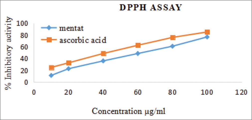
- 2,2- diphenyl-1-picrylhydrazyl free (DPPH) radical scavenging activity of Mentat tablets.
H2O2 scavenging assay
At a concentration of 100 µg/mL, the maximum % inhibition shown by Mentat tablets was (79.4%) while, at same concentration, the % inhibition of ascorbic acid was found to be (96.1%), as shown in Figure 2.
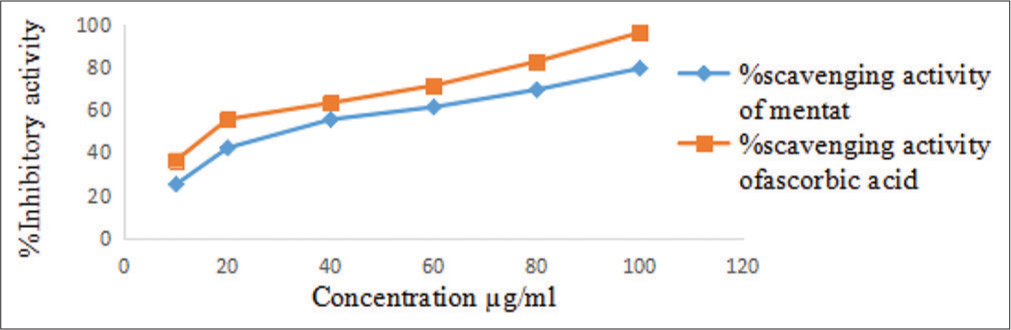
- Hydrogen peroxide scavenging activity of Mentat tablets.
In vitro anti diabetic activity of Mentat tablets
In vitro evaluation of anti-glycation activity of Mentat tablets
As shown in Table 2, the efficacy of the formulation to prevent the production of anti-glycation end (AGE) products was investigated using the anti-glycation assay on Mentat tablets. The greatest percentage of Mentat tablet inhibition was determined to be 90.09% at a dose of 1000 µg/mL, whereas the percentage of rutin standard (STD) inhibition was found to be 82.16% at the same concentration, as shown in Figure 3.
| Concentration (g/mL) | % inhibition of AGE product | |
|---|---|---|
| STD (Rutin) | Mentat tablets | |
| 100 | 61.20±0.79 | 49.66±0.58 |
| 250 | 67.86±0.21 | 47.66±0.23 |
| 500 | 73.33±0.57 | 54.83±0.39 |
| 750 | 82.9±0.32 | 63.0±0.15 |
| 1000 | 90.09±0.55 | 82.16±0.25 |
Values are expressed as mean±standard error of the mean (n=3). AGE: Anti-glycation end, STD: Standard.
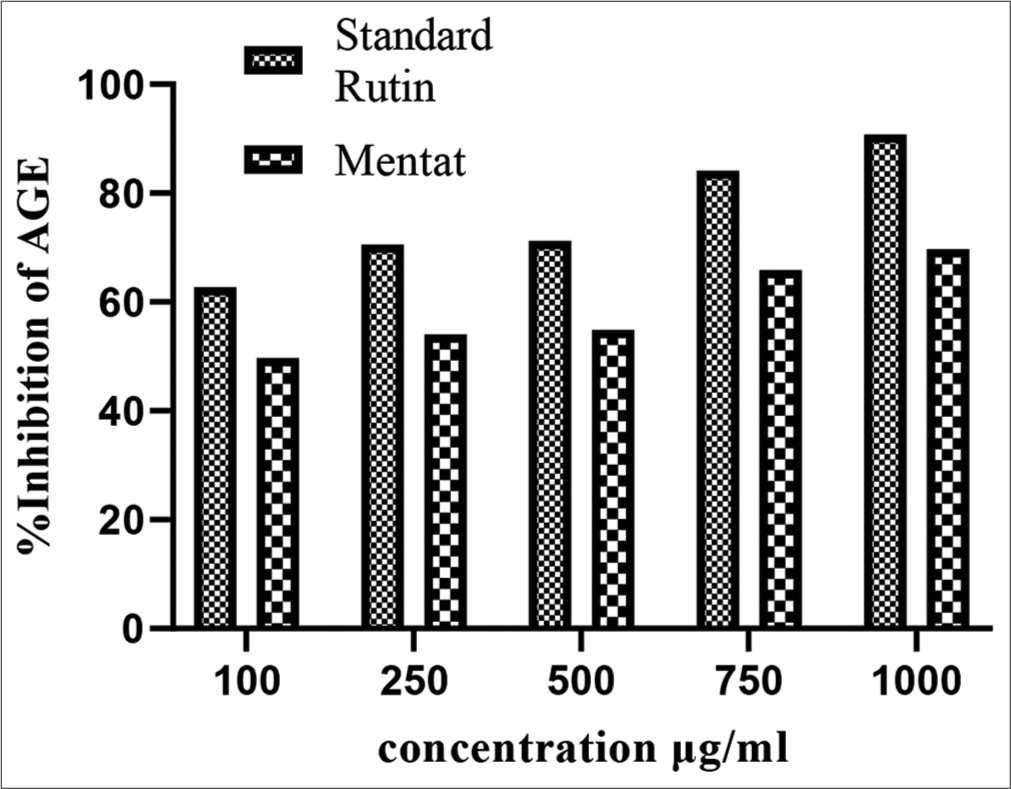
- Effect on Mentat tablets on % inhibition of anti-glycation end (AGE) products.
Determination of Insulin secretion using MIN-6 cell line
As shown in Table 3, Mentat tablets, administered at a dose of 1000 µg/mL in a glucose concentration of 11 mM, demonstrated insulin secretion. As shown in Figure 4, the highest insulin secretion was noted at 1000 µg/mL of Mentat tablets. The inclusion of Mentat tablets dramatically increased the dose-dependent release of insulin.
| Concentration (µg/mL) | Insulin secretion (ng/mL) | |
|---|---|---|
| Mentat tablets | STD | |
| 0 | 1.87±0.66 | 1.91±1.35 |
| 5 | 2.20±1.51* | 2.41±1.68** |
| 50 | 3.36±1.66* | 3.56±2.28** |
| 500 | 5.24±1.34** | 6.83±1.55*** |
| 1000 | 6.33±1.49** | 8.31±1.23*** |
The values are shown as the three observations’ mean±standard error of the mean. Comparing with base concentration, *P<0.01, **P<0.001 and ***P<0.0001. Dunnett’s test was conducted after a two-way analysis of variance analysis of the data, STD: Standard
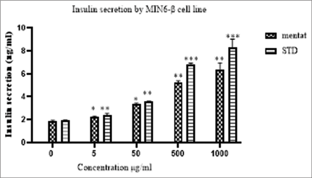
- Effect of Mentat tablets on Mouse insulinoma 6 cell line insulin secretion. *p<0.01, **p<0.001, ***p<0.0001 when compared with base concentration.
In vivo evaluation of OGTT of Mentat tablets
As shown in Figure 5 and Table 4, the Mentat tablets (100 mg/kg) and tablets (200 mg/kg) were effective in lowering high blood glucose levels in all treatment groups. Within 60 and 120 min of the test compound being administered, Mentat tablets (200 mg/kg) significantly (P < 0.001) decreased the blood glucose level.
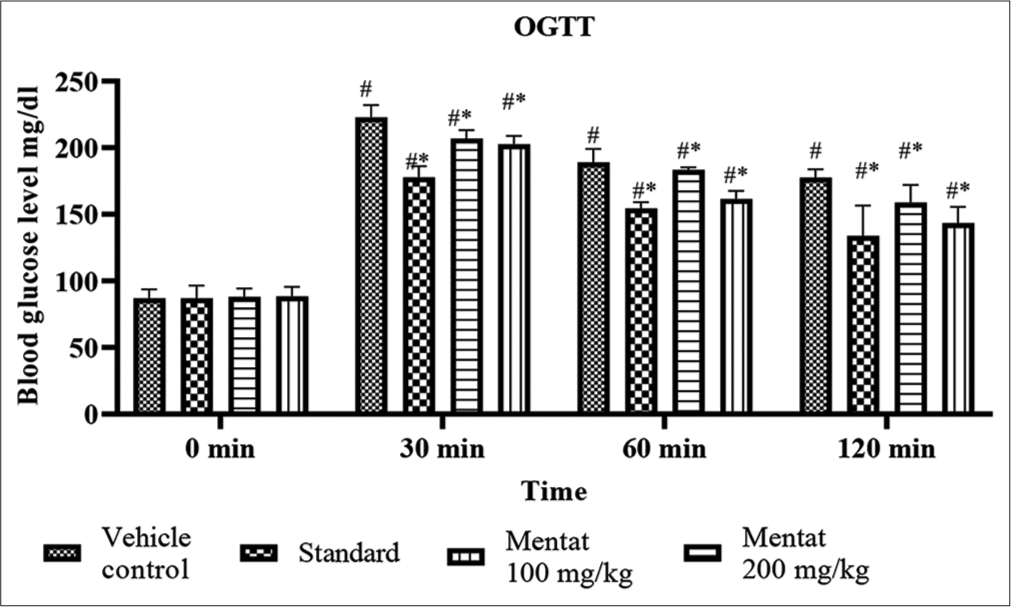
- Effect of Mentat on oral glucose tolerance test. OGTT: Oral glucose tolerance test. *p<0.05 when compared with blood concentration at 0 min; #-refers to concentration of blood glucose level at 0 min.
| S. No. | Group (n=4) | Blood glucose level (mg/dL) | |||
|---|---|---|---|---|---|
| 0 min | 30 min | 60 min | 120 min | ||
| 1. | Vehicle control | 87.0±3.366 | 222.75±4.571# | 189±4.143# | 177.5±3.225# |
| 2. | STD Glibenclamide | 86.0±4.813 | 177.75±4.210#* | 154.5±2.253#* | 133.5±2.855#* |
| 3. | Mentat tablets 100 mg/kg | 88.5±3.315 | 207±3.135#* | 183.5±2.462#* | 158.7±3.391#* |
| 4. | Mentat tablets 200 mg/kg | 88.0±3.476 | 202.5±3.227#* | 161±3.015#* | 143.2±4.230#* |
The data (n=4) were shown as mean±standard error of the mean. *P<0.0001 in the vehicle group case and #P<0.05 in the 0 min comparison case. To examine the data, two-way analysis of variance was employed. OGTT: Oral glucose tolerance test, STD: Standard.
In vivo evaluation of antidiabetic activity using HFD and STZ-induced DN in Wistar rats
As shown in Table 5, in HFD- and STZ-induced DN in Wistar rats, the treatment groups received Mentat tablets (100 mg/kg) and Mentat tablets (200 mg/kg) and were found to be effective in lowering raised blood glucose levels during 21 days. This is shown in Table 2. As depicted in Figure 6, Mentat pills (200 mg/kg) significantly (P < 0.01) reduced blood glucose levels in comparison to the illness group.
| S. No. | Group | Blood glucose level (mg/dL) | |||
|---|---|---|---|---|---|
| Day 35 | Day 42 | Day 49 | Day 56 | ||
| 1. | Vehicle | 99.75±2.495 | 100.25±3.424 | 105±4.564 | 110±2.176 |
| 2. | Disease | 335.2±1.600 | 364.75±3.81 | 372.2±5.632# | 390.5±5.952# |
| 3. | STD | 287.2±4.269 | 225±13.90 | 157.5±9.951** | 145.3±5.204** |
| 4. | Mentat tablets 100 mg/kg | 293.75±4.26 | 238.70±5.549 | 207.1±13.28* | 183.7±8.625* |
| 5. | Mentat tablets 200 mg/kg | 280±4.564 | 257.5±10.28 | 162±4.090** | 153±8.095** |
Data was analyzed by two-way ANOVA followed by Dunnett’s Test. #p< 0.0001 when compared with vehicle and *p<0.05, **p<0.01 when compared with vehicle group, STD: Standard.
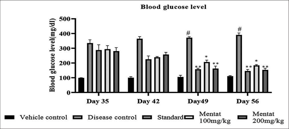
- Effect of Mentat on blood glucose level. Data was analyzed by two-way ANOVA followed by Dunnett’s Test. #p< 0.0001 when compared with vehicle and *p<0.05, **p<0.01 when compared with vehicle group.
Evaluation of Mentat tablets on lipid parameters in HFD- and STZ-induced DN in Wistar rats
As illustrated in Table 6, the treatment groups showed a significant (P < 0.01) decrease in serum levels of triglycerides (TG), total cholesterol (TC) and low-density lipoprotein cholesterol (LDL-C) in HFD- and STZ-induced diabetic rats compared to the disease control group. High-density lipoprotein cholesterol (HDL-C) was significantly (P < 0.01) increased in treatment groups compared to disease control group. Mentat (200 mg/kg) dose showed a significant (P < 0.01) decrease in serum levels of TG, TC & and LDL-C and a significant (P < 0.01) increase in HDL-C levels, as shown in Figure 7.
| S. No. | Groups | TC (mg/dL) | TG (mg/dL) | HDL-C (mg/dL) | LDL-C (mg/dL) |
|---|---|---|---|---|---|
| 1. | Vehicle control | 117±3.916 | 88.75±2.053 | 49.13±1.877 | 31.75±2.031 |
| 2. | Disease control | 210.8±4.400# | 202.2±3.483# | 23.65±1.672# | 136.96±2.154# |
| 3. | STD glibenclamide | 175.5±13.08** | 164.1±10.68** | 35.53±2.619** | 110.3±5.74** |
| 4. | Mentat tablets 100 mg/kg | 179.1±4.921* | 169.3±6.819* | 31.73±1.944* | 116.16±4.464* |
| 5. | Mentat tablets 200 mg/kg | 176.5±5.091** | 159.5±9.608** | 34.53±1.979** | 112.16±6.808** |
TC: Total cholesterol, TG: Triglycerides, HDL-C: High-density lipoprotein cholesterol, LDL-C: Low-density lipoprotein cholesterol. Values are expressed as mean ± SEM (n=6). Data was analyzed by one-way ANOVA followed by Dunnett’s Test. # p<0.0001 when compared with vehicle and* p<0.05 **p<0.01, when compared with disease group, STD: Standard.
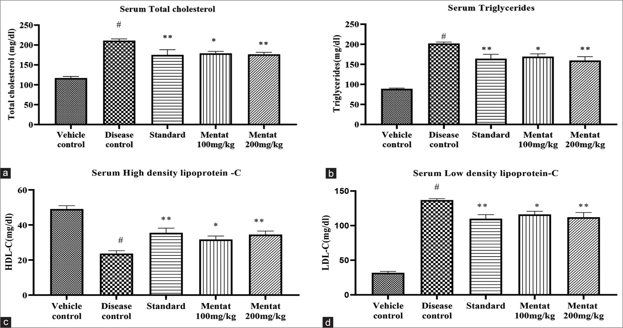
- (a) Effect of Mentat tablets on total cholesterol. (b) Effect of Mentat tablets on triglycerides. (c) Effect of Mentat tablets on high-density lipoprotein cholesterol. (d) Effect of Mentat tablets on low-density lipoprotein cholesterol. #p<0.0001 when compared to vehicle control,( **p<0.001, *p<0.01).
Evaluation of Mentat tablets on renal parameters in HFD- and STZ-induced DN in Wistar rats
According to Table 7, comparison to the disease control group, the administration of Mentat (100 mg/kg), Mentat (200 mg/kg) and glibenclamide resulted. According to Table 6 in a substantial (P < 0.01) rise in the levels of uric acid, creatinine, urea and blood urea nitrogen (BUN) in urine. The treatment groups’ urine albumin levels were considerably lower (P < 0.01) than those of the disease control group. As seen in Figure 8, Mentat (200 mg/kg) significantly decreased (P < 0.01) the level of urine albumin and significantly increased (P < 0.01) the levels of uric acid, creatinine, urea and BUN.
| S. No | Groups | Urine albumin (mg/24 h) | Urine uric acid (mg/dL) | Urine creatinine (mg/dL) | Urine urea (mg/dL) | BUN (mg/dL) |
|---|---|---|---|---|---|---|
| 1. | Vehicle Control | 22.18±2.524 | 3.736±0.047 | 54.18±1.140 | 83.68±0.861 | 39.50±3.019 |
| 2. | Disease Control | 181.4±6.371# | 1.587±0.124# | 18.77±0.346# | 14.0±1.238# | 6.50±1.056# |
| 3. | STD Glibenclamide | 129±7.98** | 2.112±0.115** | 36.63±4.97** | 30.05±3.155** | 14.02±0.577** |
| 4. | Mentat tablets 100 mg/kg | 137.8±6.725* | 1.975±0.137* | 31.77±4.522* | 25.87±5.092* | 11.83±1.108* |
| 5. | Mentat tablets 200 mg/kg | 134.3±7.135** | 2.078±0.069** | 33.95±3.383** | 28.97±2.469** | 13.5±1.765** |
BUN: Blood urea nitrogen, Values are expressed as mean ± SEM (n=6). Data was analyzed by one-way ANOVA followed by Dunnett’s Test. # p<0.0001 when compared with vehicle and *p<0.05, **p<0.01, when compared with disease group, STD: Standard.
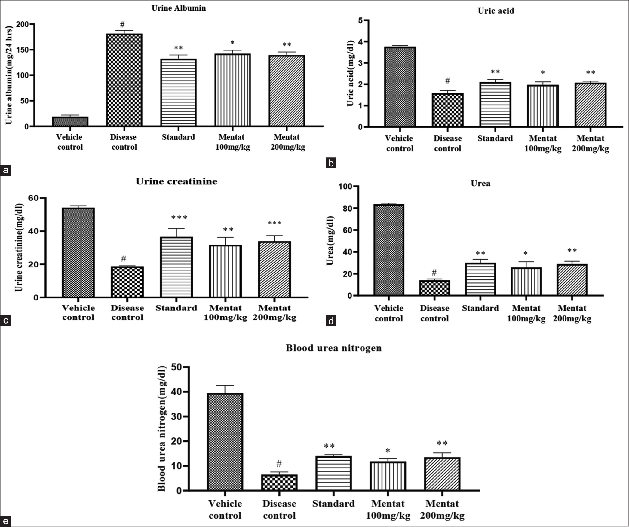
- (a) Effect of Mentat tablets on Urine albumin. (b) Effect of Mentat tablets on Uric acid. (c) Effect of Mentat tablets on urine creatinine. (d) Effect of Mentat tablets on urine urea. (e) Effect of Mentat tablets on blood urea nitrogen. #p<0.0001 when compared to vehicle control, (***p<0.0001, **p<0.001, *p<0.01).
Creatinine Evaluation of the antioxidant activity of Mentat tablets using homogenates (kidneys and pancreas)
Effect of Mentat tablets on catalase (CAT) levels
According to Table 8, tablets (100mg/kg) and Mentat tablets (200 mg/kg) showed significant (P < 0.05, P < 0.01) increases in the CAT levels in the homogenates of kidneys and pancreas in HFD and STZ induced DN when compared with disease group, as shown in Figure 9a.
| S. No. | Groups | CAT (µmol/g wt. of tissue) | |
|---|---|---|---|
| Kidney | Pancreas | ||
| 1. | Vehicle control | 71.17±2.546 | 75.07±3.125 |
| 2. | Disease control | 34.57±1.835# | 35.33±1.658# |
| 3. | STD glibenclamide | 67.35±2.326** | 57.12±1.171** |
| 4. | Mentat tablets 100 mg/kg | 50.33±1.819* | 47.17±2.753* |
| 5. | Mentat tablets 200 mg/kg | 60.33±3.135** | 62.33±4.186** |
CAT: Catalase, #p<0.0001 when compared to vehicle control, (**p<0.001, *p<0.01), STD: Standard.
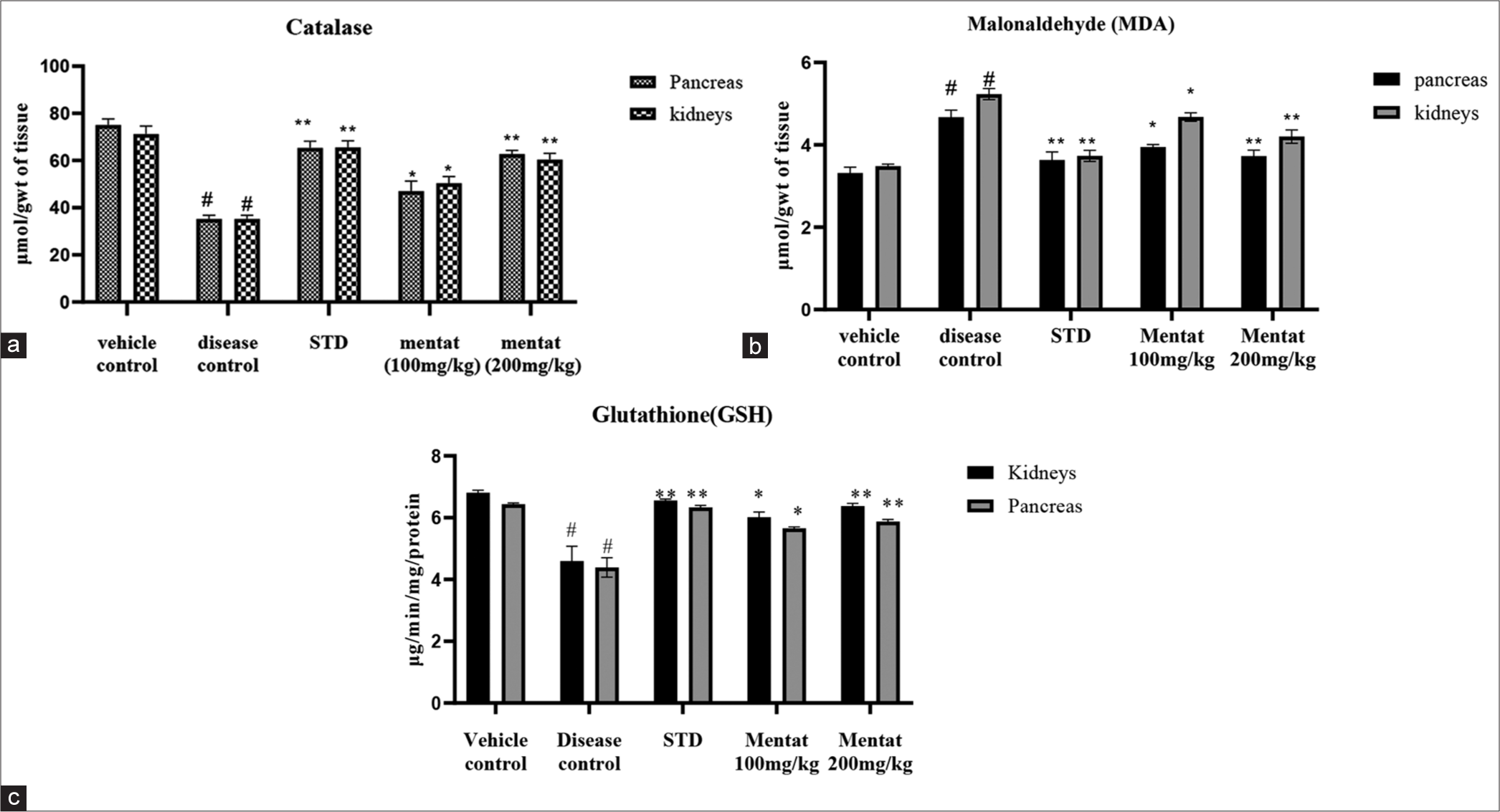
- (a) Effect of Mentat tablets on catalase. (b) Effect of Mentat tablets on malondialdehyde. (c) Effect of Mentat tablets on glutathione. #p<0.0001 when compared to vehicle control,( **p<0.001, *p<0.01). STD: Standard.
Effect of Mentat on malondialdehyde (MDA) levels
According to Table 9, (100 mg/kg) and Mentat (200 mg/kg) showed significant (P < 0.05, P < 0.01) decreases in the MDA levels in the homogenates of kidneys and pancreas in HFD- and STZ-induced DN when compared with disease group, as shown in Figure 9b.
| S. No. | Groups | MDA (µmol/g wt. of tissue) | |
|---|---|---|---|
| Kidney | Pancreas | ||
| 1. | Vehicle control | 3.317±0.058 | 3.483±0.052 |
| 2. | Disease control | 4.670±0.1255# | 5.233±0.178# |
| 3. | STD glibenclamide | 3.6±0.1016** | 3.733±0.048** |
| 4. | Mentat tablets 100 mg/kg | 3.950±0.3219* | 4.677±0.235* |
| 5. | Mentat tablets 200mg/kg | 3.727±0.256** | 4.200±0.186** |
MDA: Malondialdehyde, Values are expressed as mean ± SEM (n=6). #p<0.0001 when compared to vehicle control, (**p<0.001, *p<0.01). Data was analyzed by one-way ANOVA followed by Dunnett’s test, STD: Standard.
Effect of Mentat on glutathione (GSH) levels
According to Table 10, tablets (100 mg/kg) and Mentat tablets (200 mg/kg) showed significant (P < 0.05, P < 0.01) increases in the GSH levels in the homogenates of kidneys and pancreas in HFD- and STZ-induced DN when compared with disease group, as shown in Figure 9c.
| S. No. | Groups | Reduced GSH (µg/min/mg/protein) |
|
|---|---|---|---|
| Kidney | Pancreas | ||
| 1. | Vehicle control | 6.81±0.153 | 6.433±0.093 |
| 2. | Disease control | 4.593±0.25# | 4.390±0.054# |
| 3. | STD glibenclamide | 6.56±0.793** | 6.340±0.78** |
| 4. | Mentat tablets 100 mg/kg | 6.017±0.181* | 5.653±0.25* |
| 5. | Mentat tablets 200mg/kg | 6.377±0.235** | 5.880±0.16** |
GSH: Glutathione, Values are expressed as mean ± SEM (n=6). #p<0.0001 when compared to vehicle control, (**p<0.001, *p<0.01). Data was analyzed by one-way ANOVA followed by Dunnett’s test, STD: Standard.
Histopathology
As shown in Figures 10 and 11, the histopathological findings of kidneys revealed that β-cells were destroyed due to the induction of STZ in the HFD+STZ group. After treatment, the destruction of β-cells seemed to be repaired in the treatment group, indicating that the treatment was efficacious.
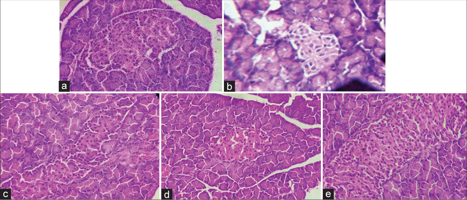
- Histopathology of pancreas in high fat diet and streptozotocin induced diabetic nephropathy in Wistar rats (a) Vehicle control, (b) disease control, (c) standard, (d) Mentat 100 mg/kg and (e) Mentat 200 mg/kg.
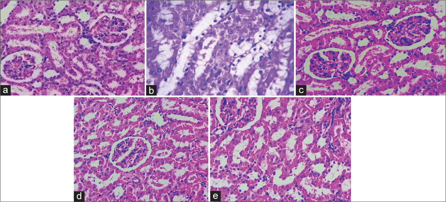
- Histopathology of kidneys in high fat diet and streptozotocin induced diabetic nephropathy in Wistar rats (a) vehicle control, (b) disease control, (c) standard, (d) Mentat 100 mg/kg and (e) Mentat 200 mg/kg.
Histopathological findings of the pancreas revealed a moderate decrease in Langerhans islets in the disease group. After treatment, there was a minimal decrease.
DISSCUSSION
A genetic or acquired defect in insulin production, or insufficient pancreatic insulin production, results in DM, a chronic condition. This deficiency causes increased blood glucose levels, which harm many body systems, including the blood vessels and neurons. DN is a clinical illness characterised by chronic albuminuria (>300 mg/24 h), high arterial blood pressure and a progressive decline in GFR. Particularly susceptible to the negative effects of STZ, a naturally occurring hormone derived from Streptomyces achromogenes, are the insulin-producing beta-cells in the pancreas of mammals. STZ-induced pancreatic injury is a common model used to simulate T2DM in mice. This damage usually manifests as traits similar to those of DN in humans.
Mentat tablets are a phytomedicinal formulation. The chief chemical constituents are brahmi, madhukaaparni, ashwagandha, jatamansi, haritaki and sunthi. The chemical constituents present in Mentat tablets are bacosides, withanolides, asiatic acid, jatamansic acid, chebulic acid and zingiberene. This medicine is used as a neuroprotective, but the constituents also possess anti-diabetic property; hence, it is repurposed as an anti-diabetic drug in the present study.
Reactive oxygen species (ROS) are crucial to the aetiology, pathophysiology and effects of diabetes. The diabetogenic effects of STZ are caused by the cytotoxicity of ROS in pancreatic β-cells, which decreases insulin production and release. The H2O2 radical scavenging and DPPH tests were used to evaluate the antioxidant activity of Mentat tablets. Because they have the ability to donate electrons and hydrogen through their hydroxyl groups, powerful antioxidants can prevent the production of free radicals caused by important components. The standard ascorbic acid concentration of 100 µg/mL was shown to have the highest antioxidant action. The % inhibition of DPPH by Mentat tablets was found to be 76.55%, and the % inhibition of H2O2 by Mentat tablets was found to be 79.55%.
The formation of anti-glycation end products (AGEs) in renal tissue is a major cause of DN. AGEs were found in almost all of the tissues examined from STZ-induced diabetic rats. Moreover, the kidneys are more likely than other organs to produce AGEs. The production of AGEs is the only mechanism responsible for the structural abnormalities observed in the kidneys of nephropathic animals. Inhibiting the AGE generation is one potential treatment for DN. The current investigation shows that Mentat tablets may have an inhibitory effect on the production of AGEs. When Mentat pills were compared to rutin as a reference, it was discovered that 82% of the AGEs were inhibited. Type 2 diabetes is a metabolic disorder characterised by the inability of beta-cells to secrete enough insulin to maintain glucose homeostasis. MIN6 cells secrete insulin in response to glucose and other secretagogues. The insulin secretion was carried out at different concentrations. The maximum insulin secretion was observed at the concentration of 1000 µg/mL when compared with the standard. Insulin secretion at 1000 µg/mL was found to be significant (P < 0.01) when compared with base concentration.
OGTT is carried out to test the body’s ability to tolerate glucose. OGTT was carried out on test formulation (Mentat tablets). Mentat tablets (100 mg/kg) and (200 mg/kg) showed a significant (P < 0.05) decrease in blood glucose level at 60 and 120 min when compared with that of vehicle.
Type 2 DM is brought on by a HFD. Since insulin resistance linked to diabetes is triggered by fat. Insulin resistance and lipotoxicity are caused by the reduction of insulin-regulated glucose metabolism brought on by the accumulation of free fatty acids in plasma. The blood glucose level was monitored during the 21 days of treatment and showed that there was a significant (P < 0.05) reduction in blood glucose level in HFD and STZ induced DN rats. The test formulation, that is, Mentat tablets (200 mg/kg), was found to be effective when compared with the base concentration of glucose followed by Mentat tablets (100 mg/kg).
Since hyperlipidaemia and diabetes are frequently linked, estimating lipid parameters is crucial. Since insulin inhibits hormone-sensitive lipase, the primary cause of the unusually high concentration of lipids in serum diabetes is the mobilisation of free fatty acids from peripheral fat stores. Therefore, the prominent hyperlipidaemia that is associated with diabetes may be thought of as the result of the fat deposit’s lipolytic hormones acting unchecked. It was found that disease control had high levels of TC, TG and LDL-C and low levels of HDL-C. Upon administration of standard, Mentat (100 mg/kg) and Mentat (200 mg/kg) significantly (P < 0.01) restored normal lipid levels by decreasing the levels of TC, TG and LDL-C and increasing the levels of HDL-C.
Insufficient blood filtration is caused by elevated blood glucose levels and injury to the blood vessels found in the kidneys. Elevated albumin levels in urine are linked to DN due to a lower rate of glomerular filtration. Therefore, the estimation of renal parameters is crucial. The disease group was found to have high amounts of albumin and low levels of creatinine, urea, uric acid and BUN in their urine. On administration of Mentat tablets in dose of 100mg/kg and 200mg/kg significantly(P<0.01) restored normal levels of urine parameters such as increase in the levels of uric acid, urea, createnine and blood urea nitrogen (BUN) while at the same time it was found that mentat tablets decreased the levels of albumin indicating effective treatment of Diabetic nephropathy. Reactive oxygen species (RNS), which comprise charged species such as superoxide and hydroxyl radical as well as uncharged species like H2O2 and singlet oxygen, are increased when oxidative stress is present. Hypothetical causal reasons for oxidative stress in diabetes include the auto-oxidation of glucose, alterations in redox equilibrium, reductions in low-molecular-weight antioxidant molecules such as GSH and Vitamin E and compromised antioxidant defence mechanisms such as superoxide dismutase and CAT. The oxidative stress parameters such as MDA, CAT and GSH assay were carried out to determine the amount and extent of oxidative stress in animals due to the presence of ROS formed from the induction of HFD and STZ. All the treatment groups showed a significant (P < 0.05, P < 0.01) decrease in the MDA and a significant (P < 0.05, P < 0.01) increase in the CAT and GSH levels in HFD- and STZ-induced DN. Histopathological studies of kidneys and pancreas show that there are no changes in the cell structure/no abnormality detected in the vehicle group. Mild to moderate cytolytic changes with vacuolisation and mild-to-moderate swelling of the tubular epithelium were observed in the disease group of pancreas and kidneys, respectively. Mentat tablets (200 mg/kg) were found to show mild recovery, while Mentat tablets (100 mg/kg) showed moderate recovery of kidneys and pancreas due to the changes that occurred by disease progression.
CONCLUSION
The present study offers pertinent proof that the phyto-medicinal formulation known as ‘Mentat tablets’ reduces the risk of DN and the metabolic abnormalities linked to DM. After assessing the Mentat pill quality control test, in vitro and in vivo activity were assessed. Since the production of ROS is linked to DN, an in vitro assessment of Mentat tablets was conducted to determine their anti-oxidant capability. It has been discovered that Mentat tablets have antioxidant properties. In addition, effective in preventing the growth of AGEs, a crucial component in the development of DN was Mentat pills. The outcomes further demonstrated that Mentat tablets, in addition to their antioxidant and antihyperglycemic activities, possess an innate potential to stimulate insulin production by the MIN6-cell line. In vivo study reveals that Mentat tablet (100 mg/kg) and (200 mg/kg) showed significant antidiabetic activity in HFD/STZ induced DN in rats. Mentat tablets (200 mg/kg) showed better results in a reduction of elevated blood glucose levels during 21 days of treatment. It may be concluded by reducing hyperglycaemia, oxidative stress and DN indicators, Mentat pills protect against kidney damage in rats with HFD/STZ-induced DN.
Ethical approval
The study/research was in accordance with New Delhi, India’s Committee for Control and Supervision of Experiments on Animals (CCSEA) regulations, the Institutional Animal Ethics Committee (IAEC), Bharati Vidyapeeth’s College of Pharmacy (BVCP) (BVCP/IAEC/25/12/2022), gave its approval to the experimental protocol.
Declaration of patient consent
Patient’s consent was not required as there are no patients in this study.
Conflicts of interest
There are no conflicts of interest.
Use of artificial intelligence (AI)-assisted technology for manuscript preparation
The authors confirm that there was no use of artificial intelligence (AI)-assisted technology for assisting in the writing or editing of the manuscript and no images were manipulated using AI.
Financial support and sponsorship
Nil.
References
- Antidiabetic and hypolipidemic activity of Helicteres isora in animal models. J Ethnopharmacol. 2002;81:343-9.
- [CrossRef] [Google Scholar]
- Spices and condiments in the management of diabetes mellitus. Res J Pharm Tech. 2011;4:37-42.
- [Google Scholar]
- Anti diabetic activity of Thuja occidentalis Linn. Res J Pharm Technol. 2008;1:362-5.
- [Google Scholar]
- Antidiabetic potential of Balarishta prepared by traditional and modern methods in alloxan induced diabetic rats. Asian J Res Chem. 2014;7:659663.
- [Google Scholar]
- Utilization pattern of anti-diabetic drugs in type 2 diabetes mellitus in tertiary care hospital. Res J Pharm Technol. 2017;10:2063-8.
- [CrossRef] [Google Scholar]
- Antidiabetic potential of aqueous and alcoholic leaf extracts of Pithecellobium dulce. Asian J Res Chem. 2009;2:83-5.
- [Google Scholar]
- Diagnosis and classification of diabetes mellitus. Diabetes Care. 2009;32(Suppl 1):S62-7.
- [CrossRef] [Google Scholar]
- Diabetes mellitus and its complications in India. Nat Rev Endocrinol. 2016;12:357-70.
- [CrossRef] [Google Scholar]
- Drug repositioning: New approaches and future prospects for life-debilitating diseases and the COVID-19 pandemic outbreak. Viruses. 2020;12:1058.
- [CrossRef] [Google Scholar]
- Hypoglycaemic and hypolipidaemic effects of Withania somnifera root and leaf extracts on alloxan-induced diabetic rats. Int J Mol Sci. 2009;10:2367-82.
- [CrossRef] [Google Scholar]
- The effect of oral administration of green tea and ginger extracts on blood glucose in diabetic rats. Int J Drug Deliv Technol. 2019;39:418-23.
- [CrossRef] [Google Scholar]
- Developing placebos for clinical research in traditional Chinese medicine: Assessing organoleptic properties of three dosage forms (oral liquid, capsule and granule) Front Pharmacol. 2021;12:673729.
- [CrossRef] [Google Scholar]
- Antioxidant and antimicrobial activities of Laetiporus sulphureus (Bull.) Murrill. Food chemistry. 2007;101:267-73.
- [CrossRef] [Google Scholar]
- Hydrogen peroxide radical scavenging and total antioxidant activity of hawthorn. Chem J. 2012;2:9-12.
- [Google Scholar]
- Beneficial role of Taurine on Biochemical parameters of diabetic female rats. Int J Pharm Qual Assur. 2020;10:662-6.
- [CrossRef] [Google Scholar]
- Antiglycation, antioxidant and toxicological potential of polyphenol extracts of alligator pepper, ginger and nutmeg from Nigeria. Asian Pac J Trop Biomed. 2012;2:727-32.
- [CrossRef] [Google Scholar]
- Pancreatic beta cell line MIN6 exhibits characteristics of glucose metabolism and glucose-stimulated insulin secretion similar to those of normal islets. Diabetologia. 1993;36:1139-45.
- [CrossRef] [Google Scholar]
- Antihyperglycemic effects of three extracts from Momordica charantia. J Ethnopharmacol. 2003;88:107-11.
- [CrossRef] [Google Scholar]
- Hypoglycemic and antidiabetic effect of ethanolic extract of leaves of Annona squamosa L. in experimental animals. J Ethnopharmacol. 2005;99:75-81.
- [CrossRef] [Google Scholar]
- Anti-diabetic potential and Indian medicinal plants. J Herb Med Toxicol. 2008;2:45-50.
- [Google Scholar]
- Treatment of mice with an herbal preparation (mentat) protects against radiation-induced mortality. Phytother Res. 2003;17:876-81.
- [CrossRef] [Google Scholar]
- Dapagliflozin ameliorates glycemic state, lipid profile and renal functions in type 2 diabetic rats. Benha Med J. 2020;37:636-52.
- [CrossRef] [Google Scholar]
- Aquilaria spp. (Agarwood) as source of health beneficial compounds: A review of traditional use, phytochemistry and pharmacology. J Ethnopharmacol. 2016;189:331-60.
- [CrossRef] [Google Scholar]
- Protective effects of curcumin on diabetic nephropathy via attenuation of kidney injury molecule 1 (KIM and neutrophil gelatinase-associated lipocalin (NGAL) expression and alleviation of oxidative stress in rats with type 1 diabetes. Iran J Basic Med Sci. 2019;22:376-83.
- [Google Scholar]


