
Translate this page into:
Honey supplemented with Vitamin C prevents dyslipidaemia and oxidative stress induced by exposure to lead acetate in Wistar rats
*Corresponding author: Olorunsola Israel Adeyomoye, Department of Physiology, University of Medical Sciences, Ondo City, Nigeria. oadeyomoye@unimed.edu.ng
-
Received: ,
Accepted: ,
How to cite this article: Adeyomoye OI, Olaniyan OT, Adewumi N, Anyakudo MM. Honey supplemented with Vitamin C prevents dyslipidaemia and oxidative stress induced by exposure to lead acetate in Wistar rats. Indian J Physiol Pharmacol 2021;65:229-36.
Abstract
Objectives:
Lead is an environmental toxicant known to cause dyslipidaemia through oxidative stress damage. The therapeutic potential of honey has widely been reported. However, there is a paucity of reports on its effects on lipid profile in Wistar rats exposed to lead.
Materials and Methods:
The effects of honey on antioxidants and lipid profile indicators were studied in 25 male Wistar rats. The rats were randomly assigned to one of the five groups (n = 5) as follows: Group I served as the control and received deionised water; Group II served as a lead acetate group (40 mg/kg bw) and Groups III, IV and V served as lead acetate groups coadministered with honey (1 mL/kg bw), Vitamin C (100 mg/kg bw) and honey+Vitamin C, respectively. The treatments were orally administered for 28 days. Body and liver weights were determined using an analytical weighing balance. Glucose and lead concentration, superoxide dismutase (SOD), total antioxidant capacity (TAC), malondialdehyde (MDA), and the lipid profile indicators were determined using spectrophotometry. The liver histology was assessed by haematoxylin and eosin staining techniques. Statistical analysis was done using analysis of variance, and the results were expressed as mean ± S.E.M. at P < 0.05.
Results:
Body weight, SOD, and TAC increased significantly in the treatment groups compared to lead acetate only. However, lead, glucose concentration, MDA, total cholesterol, triglycerides, HDL, and LDL decreased significantly in the treatment groups compared to lead acetate only. Normal histoarchitecture of the liver was seen in the treatment groups compared to lead acetate, which showed areas of inflammation.
Conclusion:
These findings imply that honey prevents dyslipidaemia which is a risk factor for metabolic diseases.
Keywords
Honey
Lipid profile
Antioxidants
Lead acetate
Dyslipidaemia

INTRODUCTION
Lead is a commonly utilised heavy metal in industries. It is chemically indicated as Pb, having an atomic number of 82 and a mass of 207.2. It is used in battery manufacturing, paints, pigments, cosmetics, ceramics, and other applications.[1] It has no biological function, and exposure to it has continued to be a widespread problem, especially in developing countries.[2] Ingestion of lead-contaminated food and water, as well as inhalation of industrial emissions and gasoline containing lead compounds, is the most common source of lead exposure.[3,4] Lead is disseminated to many organs as soon as it enters the body. The bones store 90% of total body lead, 4% in the blood, and the rest in the liver and kidneys, which both help to eliminate it.[5-7] Lead has continued to be an environmental toxicant due to its persistence in use and a rise in its level. Lead-induced oxidative stress (OS) is a complex process that produces reactive oxygen species such as hydroperoxide, singlet oxygen, and hydrogen peroxides in the body, resulting in a direct antioxidant defense system deficit in cells, which leads to tissue injury in lead-poisoned tissues and damage to the cellular biomolecules that is, lipids, proteins, and DNA.[8,9]
Honey is a natural sweetener made by either honey bees or stingless bees and is widely available across the world. Honey’s physical and chemical qualities vary based on where bees obtain raw materials from plants, as well as geographical location, meteorological conditions, and flora species.[10-12] Over 200 chemical elements are found in honey, the vast majority of which are water, carbohydrates (95–99% sugar in dry matter), fructose (32–38%) as well as disaccharides and oligosaccharides.[13] Enzymes, amino acids, minerals, organic acids, tocopherols, trace elements as well as vitamins are also found in honey.[14] Evidence has shown that honey possesses strong antioxidant activity, which helps in the prevention of many diseases associated with OS damage, some of which include diabetes mellitus, cancer, kidney disease, and cardiovascular abnormalities.[15] Scavenging assays using 1,1-diphenyl-2-picrylhydrazyl and ferric reducing antioxidant power (FRAP) have been used to assess the antioxidant activity of honey.[10,11] Honey has been shown to contain phenolic compounds, flavonoids, beta-carotene, Vitamin C, and glutathione reductase, all of which contribute to its antioxidative potential.[11] Furthermore, the donation of hydrogen ions, superoxide radical activity, and sequestration of free radicals have been proposed as possible mechanisms of action of honey.[16] In addition to its antioxidative properties, honey has been shown to have antibacterial activity by preventing the growth of pathogenic strains of Streptococcus, Escherichia coli, Pseudomonas aeruginosa, and Acinetobacter baumannii.[16,17] Honey is also useful in wound healing processes by providing substances that assist the functions of leucocytes.[18,19] It also stimulates the release of cytokines from monocytes and lymphocytes.[19] The antifungal, antiviral, antidiabetic, and anti-inflammatory potentials of honey have also been reported in the literature.[19]
Vitamin C is found only in trace amounts in almost all types of honey. Its concentration and antioxidant capacity are affected by the way honey is processed and stored.[20] Vitamin C levels in honey samples ranged from low to high. The combination of honey with other agents is considered to be a good way to boost its biological activities; therefore, supplementing natural honey with Vitamin C may help to boost its antioxidant activity. Although the beneficial effects of honey have widely been reported, its effects on lipid profile indicators in lead-exposed Wistar rats have not been fully explored. This study was designed to investigate the effects of unrefined honey on lipid profile in lead-exposed Wistar rats.
MATERIALS AND METHODS
Animals
A total of 25 male Wistar rats with an average weight of 150 ± 20 g were used in this study. The animals were kept at the University of Medical Sciences’ animal house in Ondo City. They were given 2 weeks to acclimate before the experiment. All animals were exposed to similar environmental conditions, such as ventilation, temperature (18–24°C), and lighting (12 h of light and darkness). Food (grower’s pellets) and water were freely available. Lead acetate, a chemical compound of lead in powder form, was obtained from Loba Chemical Limited, India, while unrefined honey was collected from the hive by honey bees in Nigeria. Vitamin C was purchased at Albumec Pharmaceutical Limited, Kaduna, Nigeria, and used as a standard exogenous antioxidant. Although honey is said to contain Vitamin C, its percentage concentration may not be up to the dose used in this experiment.
Ethical approval
All procedures involving animal handling and the protocol of the experiment were carried out in strict compliance with the ethics and guidelines for animal care and use in research (Institution Review Committee UNIMEDTH/ REC/14082020) and also in accordance with the guidelines provided by the Medical Association Declaration of Helsinki on ethical principles for Medical Research involving experimental animals.[21]
Animal grouping
Five groups of animals were used having five rats in each group (n = 5). Group I was given deionised water and served as the control group. Group II received 40 mg/kg bw of lead acetate according to Mahmoud et al.[22] Group III received 40 mg/kg bw of lead acetate and 1 mL/kg bw of honey according to Abdulmajeed et al.[23] Group IV received 40 mg/kg bw of lead acetate and 100 mg/kg of Vitamin C according to Li et al.[24] Group V received 40 mg/kg bw of lead acetate, 1 mL/kg bw honey, and 100 mg/kg bw of Vitamin C. All treatments were coadministered with lead acetate orally for a period of 28 days according to Abdulmajeed et al.[23]
Blood collection and analysis
After 28 days of treatment, all animals from each group were anaesthetised using ketamine (100 mg/kg) and xylazine (10 mg/kg) through the intraperitoneal route.[25] The rats were opened up through midline laparotomy and blood samples were collected through cardiac puncture and centrifuged at 3000 rpm for 3 min to obtain serum. The liver was also excised, weighed, and stored in 10% formaldehyde for histological assessment.
Determination of lead concentration, liver and body weights
Lead concentration was determined using a spectrophotometry procedure and absorbance was measured at 520 nm. A digital analytical weighing scale (Develo Scale Company, China) was used to measure the liver and body weights. The relative weight of the liver was calculated by dividing the liver weight by the body weight and multiplying by 100.
Determination of glucose concentration
Blood glucose measurement was carried out using the method of Burrin and Price.[26] The reaction mixture includes a working standard glucose solution, glucose peroxidaseoxidase reagent, and 6 N HCl. A serum sample of 0.2 mL was added and the mixture was incubated for 40 min at 35°C, thereafter, a spectrophotometer (Emclab Company, Germany) was used to measure the colour intensity at 540 nm.
Determination of total antioxidant capacity (TAC)
The TAC was determined using Rubio et al.[27] By combining buffer acetate with TPTZ solution in HCl, a workable solution of FRAP was created. After that, FeCl3 was added to the mixture. A serum sample (8 μL) and 240 μL of the working solution were combined and incubated at room temperature for 10 min. At 532 nm, the optical density of the samples was measured.
Determination of superoxide dismutase (SOD) activity
The method of Misra and Fridovich[28] for SOD measurement was essentially followed. The reaction mixture consists of bicarbonate buffer (0.05 M, pH 10.2) and adrenaline (0.03 mL). A serum sample (0.1 mL) was added to the mixture and incubated for 5 min (37°C) while the absorbance was measured at 480 nm.
Determination of malondialdehyde (MDA)
The level of MDA in serum was determined using Mateos et al.’s technique.[29] The reaction mixture consists of trichloroacetic acid (1 mL 20%) and tetramethoxypropane (0.05 mL, 10 M). A serum sample of 0.1 mL was added to the mixture and thereafter incubated in a water bath for 20 min at 100°C while the absorbance was measured at 532 nm.
Determination of cholesterol level, triglycerides, low-density lipoprotein, and high-density lipoprotein
These were all measured using commercially available colorimetric assay kits (Fortress diagnostic, Ltd., UK).[30-33]
Histological preparation of the liver tissues
The liver histology was performed using a modified version of the approach described by Alturkistani et al.[34] Tissue samples were prepared by cutting them into serial slices using a rotator microtome (Biobase, China) and then placed on albumenised slides and dried for 2 min on a warm plate. The slides were dewaxed with xylene and run through absolute alcohol for 5 min before being stained with haematoxylin and eosin and examined. For each group, photomicrographs were taken at ×400.
Statistical analysis
The findings from each study group were compared using a one-way analysis of variance and the Fisher’s LSD post hoc test. The mean and standard error of the mean was used to express the results while statistical significance was defined as P < 0.05.
RESULTS
Effects of honey supplemented with Vitamin C on body and liver weights in the control and treatment groups
There was a significant increase in final body weight (P < 0.05) compared to the initial body weight in the lead acetate group with a percentage increase of 36.41% [Figure 1]. However, the control and lead acetate groups treated with honey, Vitamin C, and both showed no significant difference (P > 0.05) in final body weights compared to their initial body weights. Furthermore, there was a significant decrease (P < 0.05) in final body weight in lead acetate treated with honey, Vitamin C, and both when compared to the lead acetate group only. The mean difference in body weights was highest in lead acetate only. There was no significant difference (P> 0.05) in absolute liver weight in lead acetate treated with honey, Vitamin C, and both when compared to lead acetate only [Figure 2].
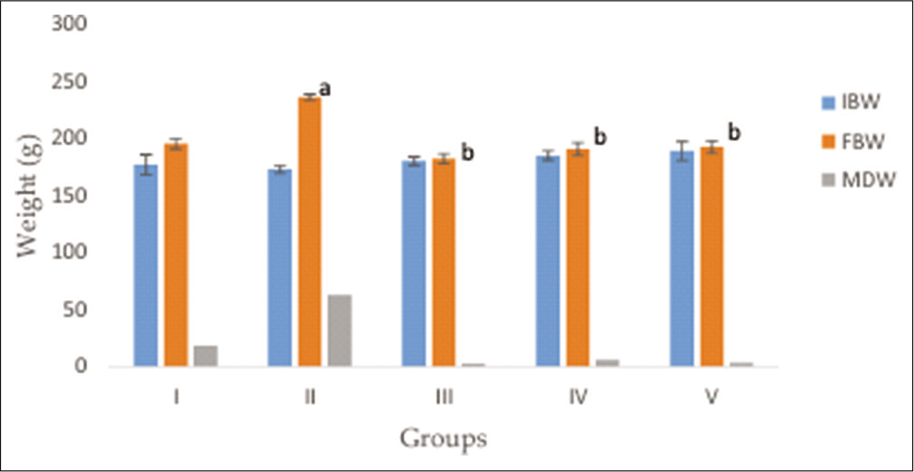
- The effects of honey supplemented with Vitamin C on body weights in the control and treatment groups. Results were expressed as mean ± SEM, P < 0.05.
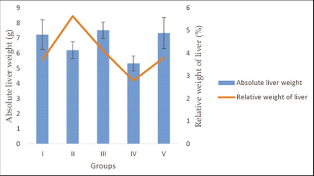
- The effects of honey supplemented with Vitamin C on absolute and relative liver weight in the control and treatment groups. Results were expressed as mean ± SEM at P < 0.05.
Effects of honey supplemented with Vitamin C on lead and glucose concentrations in the control and treatment groups
Lead concentration decreased significantly (P < 0.05) in lead acetate treated with honey, Vitamin C, and both when compared to lead acetate only [Table 1]. Glucose concentration decreased significantly (P< 0.05) in lead acetate treated with honey, Vitamin C, and both when compared to lead acetate only [Table 1]. Furthermore, glucose concentration decreased significantly (P < 0.05) in the lead acetate group treated with both honey and Vitamin C when compared to lead acetate treated with honey.
| Groups | Lead conc. (µg/dL) | Glucose conc. (mmoL) |
|---|---|---|
| I | 2.23±0.41 | 3.80±0.40 |
| II | 15.52±0.87 | 7.70±0.42 |
| III | 7.22±0.66a | 4.10±0.51a |
| IV | 9.31±1.02a | 3.63±0.31a |
| V | 7.82±0.95a | 2.64±0.44a,b |
Results were expressed as mean±SEM, P<0.05 aindicates value significantly different from lead acetate only while bindicates value significantly different from lead acetate treated with honey
Effects of honey supplemented with Vitamin C on TAC, SOD activity, and MDA in the treatment and control groups
There was a significant increase (P < 0.05) in TAC in lead acetate treated with honey, Vitamin C, and both when compared to lead acetate only [Table 2]. SOD activity increased significantly (P < 0.05) in lead acetate treated with honey, Vitamin C, and both when compared to lead acetate only [Table 2]. However, MDA was significantly lower (P < 0.05) in lead acetate treated with honey, Vitamin C, and both when compared to lead acetate only [Table 2].
| Groups | TAC (nM) | SOD (U/mL) | Malondialdehyde (μM) |
|---|---|---|---|
| I | 3.60±0.41 | 0.49±0.02 | 2.36±0.61 |
| II | 1.52±0.22 | 0.19±0.02 | 5.05±0.38 |
| III | 3.45±0.18a | 0.40±0.07a | 2.74±1.29a |
| IV | 3.69±0.27a | 0.58±0.09a | 2.51±0.45a |
| V | 3.89±0.13a | 0.70±0.08a | 2.34±0.15a |
Results were expressed as mean±SEM, P<0.05 aindicates value significantly different from lead acetate only. TAC: Total antioxidant capacity, SOD: Superoxide dismutase activity
Effects of honey supplemented with Vitamin C on lipid profile indicators in the control and treatment groups
There was a significant decrease in total cholesterol in lead acetate treated with honey and honey+Vitamin C when compared to the lead acetate group. However, there was no significant difference in total cholesterol level in the treatment groups when compared to control [Table 3]. Triglyceride level decreased significantly (P < 0.05) in the lead acetate treated with honey, Vitamin C, and both when compared to the lead acetate only. High-density lipoprotein decreased significantly (P < 0.05) in the lead acetate group treated with Vitamin C when compared to lead acetate only. Furthermore, low-density lipoprotein decreased significantly (P < 0.05) in the lead acetate group treated with honey+Vitamin C when compared to lead acetate only [Table 3].
| Groups | TC (mg/dL) | TG (mg/dL) | HDL (mg/dL) | LDL (mg/dL) |
|---|---|---|---|---|
| I | 45.95±5.16 | 31.82±8.70 | 10.24±1.19 | 31.48±4.13 |
| II | 62.89±3.84 | 51.54±4.91 | 13.82±1.04 | 33.35±4.13 |
| III | 47.68±2.00a | 23.55±1.57a | 10.18±1.47 | 25.58±1.23 |
| IV | 51.69±6.26 | 28.15±2.00a | 9.40±0.45a | 29.37±1.51 |
| V | 47.41±1.59a | 23.94±0.87a | 10.77±1.03 | 24.79±0.48a |
Results were expressed as mean±SEM, aindicates value significantly different from the Group II (lead acetate only) at P<0.05. TC: Total cholesterol, TG: Triglyceride, HDL: High-density lipoprotein, LDL: Low-density lipoprotein
Histological assessment of the hepatocytes in the control and treatment groups
The photomicrograph of the liver showed increased presence of clustered granulation on the hepatocyte in lead acetate only when compared to the control and the treatment groups. There was the loss of central venule in lead acetate only and the lead acetate group treated with honey, while the lead acetate group with honey+Vitamin C showed distortion and steatosis of the central venule [Figure 3].
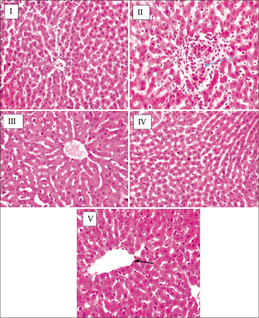
- The photomicrograph of the liver. Group I (control) and IV (lead acetate 40 mg/kg+Vitamin C 100 mg/kg) showed normal architecture of the hepatocyte, Group II (lead acetate 40 mg/ kg) showed clustered granulation of the hepatocyte (blue arrow), Group III (lead acetate 40 mg/kg+ honey 1 mL) showed distortion of central venule, Group V (lead acetate 40 mg/kg+ Vitamin C 100 mg/kg + honey 1 mL) showed steatosis and distortion of the central venule (black arrow) (H&E ×400).
DISCUSSION
Dyslipidaemia is an important risk factor for metabolic diseases such as obesity, coronary heart disease, stroke, diabetes, and many others.[35] To maintain a healthy lifestyle and prevent the morbidity associated with dyslipidaemia, it is important to have a favourable lipid profile. This study investigated the effects of honey supplemented with Vitamin C on lipid profile indicators in Wistar rats exposed to lead acetate.
Results showed a significant decrease in final body weight in lead acetate treated with honey, Vitamin C and both compared to the lead acetate group [Figure 1]. This observation is in accordance with the report of Nemoseck et al.[36] who reported that honey promotes lower weight gain than sucrose and other common sweeteners in rats. In addition, Yaghoobi et al.[37] also reported a weight decrease in overweight patients treated with natural honey. This effect of honey supplemented with Vitamin C may be due to its ability to reduce food intake through kisspeptin inhibition which enhances insulin secretion for glucose uptake.[38] However, further studies are needed to elucidate the mechanisms involved. The initial body weight increases across the groups though this increase was not significant. This increase may be because the animals do not have the same starting weights. Surprisingly, no significant difference in liver weight was seen in all the groups, implying that the liver was unaffected and, therefore, lead acetate may have had a reduced impact on the liver and exerted most of its effects through extrahepatic organs such as the spleen, heart, and kidney [Figure 2].
A significant decrease in blood glucose was observed in the lead acetate treated with honey, Vitamin C, and both. This observation supports the work of Bobiş et al.[39] who reported the hypoglycaemic effects of honey and described its use in preventing and treating different types of diabetes. He described honey as a novel antidiabetic agent that could be useful in the treatment of diabetes and its complications, as well as the potential consequences and future perspective of using honey as an antidiabetic agent. However, this finding is in contrast with Rashid et al.[40] who reported that honey had no effect on impaired glucose tolerance. Furthermore, Whitfield et al.[41] also investigated the effects of cinnamon, chromium, and magnesium-formulated honey on glycaemic control with no significant reduction in plasma glucose. Therefore, more studies are needed to ascertain the hypoglycaemic properties of honey. A significant decrease in glucose concentration was also observed in lead acetate treated with honey and Vitamin C compared to lead acetate treated with honey. This observation is in contrast with the report of Al-Obaidi et al.[42] who reported an increase in blood glucose following the administration of Vitamin C in SARS-CoV-2 patients. The observed decrease in glucose concentration may be due to the ability of the Vitamin C supplement to reduce the formation of glycated haemoglobin, which is an indicator of high blood sugar.[43]
A significant increase in TAC and SOD was observed in lead acetate treated with honey, Vitamin C, and both against lead acetate only. This increase is in support of Wang et al.[44] who reported that vitex honey had antioxidative effects on paracetamol-induced liver damage in mice. Zhao et al.[45] also reported the antioxidative effect of Apis cerana honey on alcohol-induced liver damage in mice. The increase in TAC may be due to the ability of honey to protect the blood, cells, tissues, and organs from free radical damage, which is commonly thought to be a sign of OS, generated by an imbalance in pro-oxidant and antioxidant defense systems [Table 2].[46] SOD, a metalloenzyme found in all living species’ cells, breaks down potentially hazardous oxygen molecules (ROS), preventing tissue damage as well. This enzyme carries out the catalysis of the reaction of the superoxide radical (O2) splitting into molecular oxygen (O2) and hydrogen peroxide (H2O2).[47] The significant increase in SOD in lead acetate treated with honey, Vitamin C both implies that they have the ability to potentiate SOD to eliminate scavenging radicals. Lipid peroxidation occurs when free radicals, primarily hydroxyl and hydroperoxyl radicals, damage lipid-containing compounds such as polyunsaturated fatty acids. Many cellular diseases are associated with these assaults, including cancer, neurological disease, cardiovascular abnormalities, and many others.[48] The rise in MDA levels in the lead acetate group could be a sign of OS damage [Table 2]. However, treatment with honey, Vitamin C, and both prevent MDA levels from rising. This observation supports the findings of Cheng et al [49] who reported a decrease in MDA following the treatment of carbon tetrachloride-induced liver and DNA damage mice with buckwheat honey produced in China. Therefore, honey may be acting, possibly by serving as an antioxidant to mop up free radicals generated through lead exposure.[50]
There are controversial reports in the literature on the effects of honey on lipid profiles.[36,51,52] We found a significant decrease in cholesterol in lead acetate treated with honey and honey+Vitamin C, a significant decrease in triglycerides in lead acetate treated with honey, Vitamin C, and both, a significant decrease in HDL in lead acetate treated with Vitamin C and a significant decrease in LDL in lead acetate treated with honey+Vitamin C compared to lead acetate only [Table 3]. This observation is in line with the work of Yaghoobi et al.[37] and Rasad et al.[53] who reported a decrease in cholesterol, triglycerides, and LDL in healthy subjects. The decrease in TC, TG, and LDL in Vitamin C supplemented honey may be due to its ability to block the enzyme 3-hydroxy-3-methylglutaryl coenzyme A (HMGCoA reductase), which is involved in cholesterol production.[54] It could also possibly be because of its capacity to activate LDL receptors, which aid in the clearance of cholesterol low-density lipoprotein from the circulation.[55] The decrease in HDL caused by Vitamin C supports the work of ContrerasDuarte[52] who showed that Vitamin C has the capacity to cause HDL remodelling.
Honey has widely been reported to be an anti-inflammatory agent. Hence, the evenly dispersed granulation observed in photomicrographs of the lead acetate groups treated with honey supplemented with C supports its involvement in controlling inflammatory processes [Figure 3 iii-v].[56] Honey has also been proven to decrease inflammatory mediators such as prostaglandin E2 and cyclooxygenase 2, therefore, it may be operating through these mechanisms to lower inflammation.[13] The distortion in the central venules observed in lead acetate treated with honey and honey+Vitamin C could be due to the toxic effect of lead on the vessels responsible for draining blood away from the hepatocytes.
CONCLUSION
Oral administration of honey supplemented with Vitamin C in lead acetate rats decreased the final body weight, glucose concentration, MDA, total cholesterol, triglycerides, and low-density lipoproteins while TAC and SOD activity increased. These findings support the existing literature that honey helps to prevent dyslipidaemia, a significant risk factor for many metabolic diseases, by increasing antioxidant enzyme activity and modulating lipid profile indicators [Figure 4]. Furthermore, this study showed that Vitamin C supplementation reduced HDL, the good cholesterol that has been reported to lower the risk of obesity, cardiovascular abnormalities, and other types of metabolic diseases. The reason for this effect remains unknown. Although the presence of hepatocyte granulation was used to indicate inflammation, the inability to assess other inflammatory markers is a major limitation to this study.
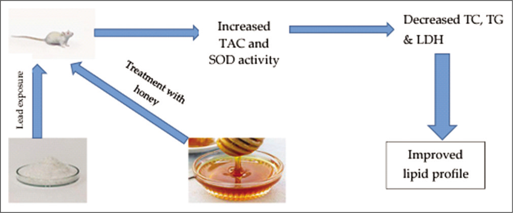
- Schematic diagram showing possible pathway of action of honey supplemented with Vitamin C in preventing dyslipidaemia in Wistar rats. TAC: Total antioxidant capacity, SOD: Superoxide dismutase, CH: Cholesterol, TG: Triglyceride, LDL: Low-density lipoprotein.
Declaration of patient consent
Patient’s consent not required as there are no patients in this study.
Financial support and sponsorship
Nil.
Conflicts of interest
There are no conflicts of interest.
References
- Lead exposure causes alteration of hematological indices in adult female Wistar rats. Ajprd. 2019;7:30-4.
- [CrossRef] [Google Scholar]
- Toxicity of lead: A review with recent updates. Interdiscip Toxicol. 2012;5:47-58.
- [CrossRef] [PubMed] [Google Scholar]
- The detrimental effects of lead on human and animal health. Vet World. 2016;9:660-71.
- [CrossRef] [PubMed] [Google Scholar]
- Lead exposure induces Alzheimers's disease (AD)-like pathology and disturbes cholesterol metabolism in the young rat brain. Toxicol Lett. 2018;296:173-83.
- [CrossRef] [PubMed] [Google Scholar]
- Role of essential trace minerals on the absorption of heavy metals with special reference to lead. Indian J Clin Biochem. 2003;18:154-60.
- [CrossRef] [PubMed] [Google Scholar]
- Genotoxic effects of lead: An updated review. Environ Int. 2010;36:623-36.
- [CrossRef] [PubMed] [Google Scholar]
- Oxidative stress in lead and cadmium toxicity and its amelioration. Vet Med Int. 2011;2011:457327.
- [CrossRef] [PubMed] [Google Scholar]
- Free radicals, metals and antioxidants in oxidative stress-induced cancer. Chem Biol Interact. 2006;160:1-40.
- [CrossRef] [PubMed] [Google Scholar]
- Antioxidant and oxidative stress: A mutual interplay in age-related diseases. Front Pharmacol. 2018;9:1162.
- [CrossRef] [PubMed] [Google Scholar]
- Endogenous and exogenous antioxidants as a tool to ameliorate male infertility induced by reactive oxygen species. Antioxid Redox Signal. 2020;33:11.
- [CrossRef] [PubMed] [Google Scholar]
- Honey and health: A review of recent clinical research. Pharmacognosy Res. 2017;9:121-7.
- [Google Scholar]
- Honey as a potential natural antioxidant medicine: An insight into its molecular mechanisms of action. Oxid Med Cell Longev. 2018;2018:8367846.
- [CrossRef] [PubMed] [Google Scholar]
- Antioxidant capacity of honey from the urban apiary: A comparison with honey from the rural apiary. Sci Rep. 2021;11:9695.
- [CrossRef] [PubMed] [Google Scholar]
- Honey: Its medicinal property and antibacterial activity. Asian Pac J Trop Biomed. 2011;1:154-60.
- [CrossRef] [Google Scholar]
- Honey: A sweet solution to the growing problem of antimicrobial resistance? Future Microbiol. 2013;8:1419-29.
- [CrossRef] [PubMed] [Google Scholar]
- Honey exposure stimulates wound repair of human dermal fibroblasts. Burns Trauma. 2013;1:32-8.
- [CrossRef] [PubMed] [Google Scholar]
- The Composition and biological activity of honey: A focus on manuka honey. Foods. 2014;3:420-32.
- [CrossRef] [PubMed] [Google Scholar]
- Potential of terpenoids and flavonoids from asteraceae as anti-inflammatory, antitumor, and antiparasitic agents. Evid Based Complement Alternat Med. 2017;2017:6196198.
- [CrossRef] [PubMed] [Google Scholar]
- RP-HPLC determination of water-soluble vitamins in honey. Talanta. 2011;83:924-9.
- [CrossRef] [PubMed] [Google Scholar]
- Guide for the Care and Use of Laboratory Animals (8th ed). Washington, DC: National Academies Press; 2011.
- [Google Scholar]
- Effects of L-cysteine on lead acetate induced neurotoxicity in albino mice. Biotech Histochem. 2016;91:327-32.
- [CrossRef] [PubMed] [Google Scholar]
- Honey prevents neurobehavioural deficit and oxidative stress induced by lead acetate exposure in male Wistar rats-a preliminary study. Metab Brain Dis. 2016;31:37-44.
- [CrossRef] [PubMed] [Google Scholar]
- Effect of vitamin C and E on antioxidative enzyme, NOS activity and NO contents in hippocampus of rats with lead poisoning. Zhejiang Da Xue Xue Bao Yi Xue Ban. 2008;37:189-92.
- [Google Scholar]
- Pharmacokinetics of ketamine and xylazine in young and old Sprague-Dawley rats. J Am Assoc Lab Anim Sci. 2013;52:567-70.
- [Google Scholar]
- Measurement of blood glucose. Ann Clin Biochem. 1985;22:327-42.
- [CrossRef] [PubMed] [Google Scholar]
- Spectrophotometric assays for total antioxidant capacity (TAC) in dog serum: An update. BMC Vet Res. 2016;12:166.
- [CrossRef] [PubMed] [Google Scholar]
- The role of superoxide anion in the autoxidation of epinephrine and a simple assay for superoxide dismutase. J Biol Chem. 1972;247:3170-5.
- [CrossRef] [Google Scholar]
- Determination of malondialdehyde (MDA) by high-performance liquid chromatography in serum and liver as a biomarker for oxidative stress. Application to a rat model for hypercholesterolemia and evaluation of the effect of diets rich in phenolic antioxidants from fruits. J Chromatogr B Analyt Technol Biomed Life Sci. 2005;827:76-82.
- [CrossRef] [PubMed] [Google Scholar]
- Direct determination of total serum cholesterol by use of double-wavelength spectrophotometry. Clin Chem. 1975;21:703-7.
- [CrossRef] [PubMed] [Google Scholar]
- A method for the sequential colorimetric determination of serum triglycerides and cholesterol. Clin Biochem. 1987;20:167-72.
- [CrossRef] [Google Scholar]
- High density lipoproteins: Measurement techniques and potential biomarkers of cardiovascular risk. BBA Clin. 2015;3:175-88.
- [CrossRef] [PubMed] [Google Scholar]
- Spectrophotometry and ultracentrifugation for measurement of plasma lipids in dogs with diabetes mellitus. J Vet Intern Med. 2018;32:93-8.
- [CrossRef] [PubMed] [Google Scholar]
- Histological stains: A literature review and case study. Glob J Health Sci. 2015;8:72-9.
- [CrossRef] [PubMed] [Google Scholar]
- Honey promotes lower weight gain, adiposity, and triglycerides than sucrose in rats. Nutr Res. 2011;31:55-60.
- [CrossRef] [PubMed] [Google Scholar]
- Natural honey and cardiovascular risk factors; effects on blood glucose, cholesterol, triacylglycerole, CRP, and body weight compared with sucrose. ScientificWorldJournal. 2008;8:463-9.
- [CrossRef] [PubMed] [Google Scholar]
- Does kisspeptin regulate food intake in humans? An important journey of discovery has begun. J Clin Endocrinol Metab. 2021;106:e1031-3.
- [CrossRef] [PubMed] [Google Scholar]
- Honey and diabetes: The importance of natural simple sugars in diet for preventing and treating different type of diabetes. Oxid Med Cell Longev. 2018;2018:4757893.
- [CrossRef] [PubMed] [Google Scholar]
- The effect of kelulut honey on fasting blood glucose and metabolic parameters in patients with impaired fasting glucose. J Nutr Metab. 2019;2019:3176018.
- [CrossRef] [PubMed] [Google Scholar]
- The effect of a cinnamon-, chromium-and magnesium-formulated honey on glycaemic control, weight loss and lipid parameters in Type 2 diabetes: An open-label cross-over randomised controlled trial. Eur J Nutr. 2016;55:1123-31.
- [CrossRef] [PubMed] [Google Scholar]
- The influence of Vitamin-C intake on blood glucose measurements in COVID-19 pandemic. J Infect Dev Ctries. 2021;15:209-13.
- [CrossRef] [PubMed] [Google Scholar]
- Impact of Rutin and Vitamin C combination on oxidative stress and glycemic control in patients with Type 2 diabetes. Clin Nutr ESPEN. 2020;35:128-35.
- [CrossRef] [PubMed] [Google Scholar]
- Antioxidant and hepatoprotective activity of vitex honey against paracetamol induced liver damage in mice. Food Funct. 2015;6:2339-49.
- [CrossRef] [PubMed] [Google Scholar]
- Antioxidant and hepatoprotective effects of A cerana honey against acute alcohol-induced liver damage in mice. Food Res Int. 2017;101:35-44.
- [CrossRef] [PubMed] [Google Scholar]
- Total antioxidant capacity-relevance, methods and clinical implications. Andrologia. 2021;53:e13624.
- [CrossRef] [Google Scholar]
- Therapeutic potentials of superoxide dismutase. Int J Health Sci (Qassim). 2018;12:88-93.
- [Google Scholar]
- Lipid peroxidation products in human health and disease. Oxid Med Cell Longev. 2013;2013:583438.
- [CrossRef] [PubMed] [Google Scholar]
- Buckwheat honey attenuates carbon tetrachloride-induced liver and DNA damage in mice. Evid Based Complement Alternat Med. 2015;2015:987385.
- [CrossRef] [PubMed] [Google Scholar]
- Protective effects of gelam honey against oxidative damage in young and aged rats. Oxid Med Cell Longev. 2014;2014:673628.
- [CrossRef] [PubMed] [Google Scholar]
- The long-term effects of feeding honey compared with sucrose and a sugar-free diet on weight gain, lipid profiles, and DEXA measurements in rats. J Food Sci. 2008;73:H1-7.
- [CrossRef] [PubMed] [Google Scholar]
- Attenuation of atherogenic apo B-48-dependent hyperlipidemia and high density lipoprotein remodeling induced by Vitamin C and E combination and their beneficial effect on lethal ischemic heart disease in mice. Biol Res. 2018;51:34.
- [CrossRef] [PubMed] [Google Scholar]
- The effect of honey consumption compared with sucrose on lipid profile in young healthy subjects (randomized clinical trial) Clin Nutr ESPEN. 2018;26:8-12.
- [CrossRef] [PubMed] [Google Scholar]
- The 3-hydroxy-3-methylglutaryl coenzyme-a (HMG-CoA) reductases. Genome Biol. 2004;5:248.
- [CrossRef] [PubMed] [Google Scholar]
- The LDL receptor. Arterioscler Thromb Vasc Biol. 2009;29:431-8.
- [CrossRef] [PubMed] [Google Scholar]
- Honey and its nutritional and anti-inflammatory value. BMC Complement Med Ther. 2021;21:30.
- [CrossRef] [PubMed] [Google Scholar]


